Ribs
The human body has twelve pairs of ribs (lat. Costae). They have a curved and rod-like shape. The ribs run bent from the thoracic vertebrae to the sternum and form a cross connection. The connection between sternum and ribs is formed by the rib cartilage. The ribs represent an important part of the bony thorax.
Anatomy
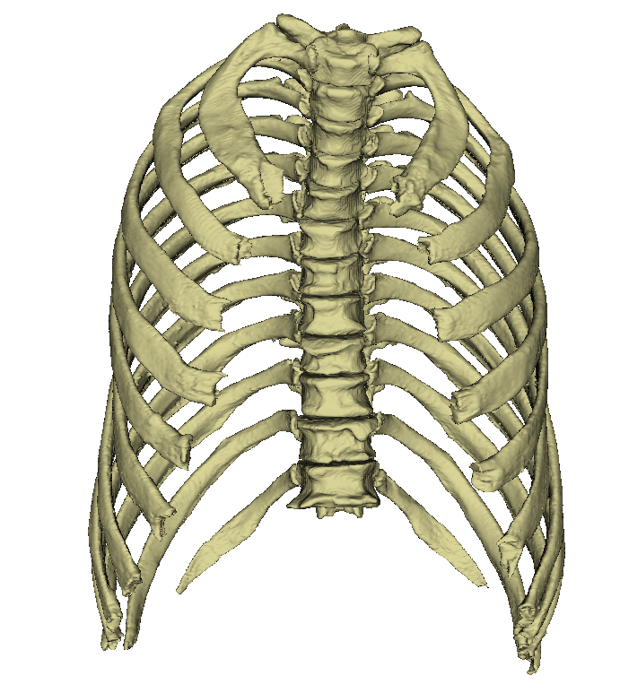
Ribs are the paired, curved, rod-shaped bones that originate on the dorsal side of the thoracic spine.
Most ribs then reach the sternum in a semicircle.
In this way, they create a cross connection between the two structures and thus serve the stability and elasticity of the thorax.
Usually the female chest is narrower and shorter than the male.
The human body has a total of twelve pairs of ribs, which corresponds to the number of thoracic vertebrae.
Each rib consists of the actual rib bone (os costale) and an adjacent section of cartilage, the costal cartilage (cartilago costalis). The individual ribs are not directly adjacent to each other, but are separated by the intercostal space. This space is partially filled by the intercostal muscles.
The ribs are connected to the spinal column by the rib heads. Most ribs touch two consecutive vertebrae. The joint surface of the head of the rib (facies articularis capitis costae) is therefore divided in two in this case.
At the neck of the rib, the rib hump (tuberculum costae) is located, which is connected to the transverse process of the thoracic vertebra at the same level via a small joint surface (facies articularis tuberculi costae). Not far from the rib neck is the angulus costae (rib angle). The flat body of the rib has a convex outer surface (facies externa) and a concave inner surface (facies interna).
The anterior part of the ribs consists of cartilage. This elastically connects the ribs to the sternum. Without this freedom of movement, the ribs could not move when breathing in and out.
Classification of the ribs

The ribs can be divided into three groups. The decisive factor here is the connection to the sternum.
Die ersten sieben Rippenpaare erreichen als sogenannte echte Rippen (Costae verae) das Brustbein direkt über ihren zugehörigen Rippenknorpel.
The next five ribs are called false rib pairs (costae spuriae). The 8th, 9th and 10th ribs are only indirectly connected to the sternum. The respective nearest rib cartilages connect and are thus involved in the construction of the costal arch (arcus costalis).
The last two " false" pairs of ribs (costae fluctuantes) of the 11th and 12th ribs usually end up freely between the muscles of the lateral abdominal wall.
Different shapes of ribs


Each pair of ribs is bilaterally symmetrical, but the shape is different in each segment. The neck of the rib extends from the head of the rib (caput costae) to the tubercle (tuberculum costae) and is located at the dorsal end of the rib. With the exception of the 1st rib, it has an upward pointing groin (crista colli costa). Lateral from the rib hump the rib body (corpus costae) bends forward forming the rib angle (angulus costae). The rib bodies of the 2nd-11th rib have irregular curvatures, the so-called surface and edge curvatures.
Due to their additional rotation around the longitudinal axis, the outer surfaces of the ribs are slightly caudal at their vertebral end and slightly cranial at their ventral end. Normally, the 1st and 12th ribs are shortest, and the 7th rib is longest. The cartilage of the rib increases in length from the 1st-7th rib, and becomes shorter again from the 8th rib. With the exception of the 1st, 11th and 12th ribs, each rib has a groove (sulcus costae) on its lower surface, in which the intercostal vessels and nerves are relatively protected.
Function
The ribs, together with the thoracic vertebrae, the costal cartilage and the sternum, form the bony ribcage. Their main task is to stabilise and protect the internal organs, especially the heart and lungs.
Movement
The ribs perform a significant movement during breathing. These are performed by the rib-vertebral joints and rib-chest joints. The lifting and lowering during inhalation and exhalation is caused by the intercostal muscles. The movement includes a frame of about 2 cm.
Develpoment
The ribs are made of somites. The bone clips ossify towards the end of the second embryonic month. The ossification starts in some ossification nuclei at the angulus costae. The bone braces do not harden completely, so adult people also have rib cartilage.