Wirbelsäule/en: Unterschied zwischen den Versionen
(Die Seite wurde neu angelegt: „Deformations of the spine can cause wear and tear and muscle tension. In principle, there are three types of deformation: Scoliosis, kyphosis and lordosis. The…“) |
Becher (Diskussion | Beiträge) |
||
| (71 dazwischenliegende Versionen von 3 Benutzern werden nicht angezeigt) | |||
| Zeile 1: | Zeile 1: | ||
| − | + | The bony spine (lat. Columna vertebralis) usually consists of 32 to 34 individual vertebrae. The [[Special:MyLanguage/Rumpf|trunk]] is carried by the spinal column. When viewed from the side, its double "s" shape becomes visible. The spinal column is divided into four sections: [[Special:MyLanguage/Halswirbelsäule|cervical spine]], [[Special:MyLanguage/Brustwirbelsäule|thoracic spine]], [[Special:MyLanguage/Lendenwirbelsäule|lumbar spine]] and [[Special:MyLanguage/Kreuz- und Steißbein|sacral bone and tailbone]]. The cervical, thoracic and lumbar vertebrae belong to the mobile vertebrae, these are connected to each other by intervertebral discs. | |
| − | The bony spine (lat. Columna vertebralis) usually consists of 32 to 34 individual vertebrae. The [[Special:MyLanguage/Rumpf| | + | |
| − | + | {{ArticleMenu_en|Links Übungsaufgaben=[[Special:MyLanguage/Übungsaufgaben: Rumpf|Trunk]][[Special:MyLanguage/Übungsaufgabe: Wirbelsäule ventral|Spine]]| | |
| − | + | Segmentereinbettung=<segmenter-embedding public wsemb-id="WirbelsaeuleMann" file="WirbelsaeuleMann.seg" height="300" width="400"/>| | |
| − | + | Links Benachbarte Strukturen=[[Special:MyLanguage/Aufbau Wirbel|Structure vertebrae]][[Special:MyLanguage/Halswirbelsäule|Cervical spine]][[Special:MyLanguage/Brustwirbelsäule|Thoracic spine]][[Special:MyLanguage/Kreuz- und Steißbein|Sacrum]]| | |
| − | + | Links Körperregionen=[[Special:MyLanguage/Knochen Rumpf|Bones trunk]][[Special:MyLanguage/Rumpf|Trunk]]| | |
| − | + | Links Organsystem=[[Special:MyLanguage/Unregelmäßige Knochen|Irregular bones]][[Special:MyLanguage/Knochen|Bones]][[Special:MyLanguage/Passiver Bewegungsapparat|Passive movement apparatus]][[Special:MyLanguage/Bewegungsapparat|Movement apparatus]]}} | |
| − | + | ||
| − | + | ||
| − | |||
| − | |||
| − | |||
| − | |||
| − | |||
| − | |||
| − | |||
---- | ---- | ||
==Anatomy== | ==Anatomy== | ||
| − | ===Sections of the spine== | + | ===Sections of the spine=== |
| − | + | {{ArticleGallery| | |
| − | + | Bild 1=<lightbox-embedding src="file:WirbelsäuleAnsicht1.png" group="image-group-1" caption="Anatomy of the spine" width="400" height="300" style=""/>| | |
| − | <lightbox-embedding src="file:WirbelsäuleAnsicht1.png" group="image-group-1" caption="Anatomy of the spine" width="400" height="300" style=" | + | Weitere Bilder=<lightbox-embedding src="file:Wirbelsäule_Ansichten2.png" group="image-group-1" caption="Anatomy of the spine"/> <lightbox-embedding src="file:Wirbelsäule_Ansichten3.png" group="image-group-1" caption="Anatomy of the spine lateral"/> |
| − | + | |title=Anatomy of the spine}} | |
| − | |||
| − | <lightbox-embedding src="file:Wirbelsäule_Ansichten2.png" group="image-group-1" caption="Anatomy of the spine"/> <lightbox-embedding src="file:Wirbelsäule_Ansichten3.png" group="image-group-1" caption="Anatomy of the spine"/> | ||
| − | |||
| − | |||
| − | Anatomy of the spine | ||
| − | |||
| − | + | {{ArticleGallery| | |
| − | + | Bild 1=<lightbox-embedding src="file:Abschnitte Wirbelsäule beschriftet.png" group="image-group-1" caption="Curvatures and sections of the spine" width="400" height="300" style=""/>| | |
| − | <lightbox-embedding src="file:Abschnitte Wirbelsäule beschriftet.png" group="image-group-1" caption="Curvatures and sections of the spine" width="400" height="300" style=" | + | Weitere Bilder= |
| − | + | |title=Curvatures and sections of the spine}} | |
| − | |||
| − | Curvatures and sections of the spine | ||
| − | |||
| − | The latin term for the spinal column is Columna vertebralis. It runs from the head to the [[Special:MyLanguage/ | + | The latin term for the spinal column is Columna vertebralis. It runs from the head to the [[Special:MyLanguage/Beckengürtel|Pelvis]], so from dorsal it represents the vertical axis of the human being. The spine of an adult human being is divided into four sections: Cervical spine (HWS), thoracic spine (BWS), lumbar spine (LWS) and sacral spine (Os sacrum). Accordingly, it also has four typical curvatures in the sagittal plane in humans. Each section is made up of individual vertebral bodies: |
*7 Cervical vertebrae (Vertebrae cervicales / C1-C7) | *7 Cervical vertebrae (Vertebrae cervicales / C1-C7) | ||
| Zeile 45: | Zeile 29: | ||
*5 Sacral vertebrae (Vertebrae sacrales / S1-S5 and Vertebrae coccygeae / Co1-Co5) | *5 Sacral vertebrae (Vertebrae sacrales / S1-S5 and Vertebrae coccygeae / Co1-Co5) | ||
| − | The different vertebrae each show a different [[Special:MyLanguage/Aufbau Wirbel| | + | The different vertebrae each show a different [[Special:MyLanguage/Aufbau Wirbel|structure of a vertebra]]. The vertebrae of the cervical, thoracic and lumbar spine are freely movable and connected to each other by the intervertebral discs. The sacral vertebrae, on the other hand, merge into the sacrum and coccyx in the course of life. |
===Shape of the spine=== | ===Shape of the spine=== | ||
| Zeile 51: | Zeile 35: | ||
Seen from the side, the double "s" curvature of the spine is visible. The four sections thus each show a characteristic curvature: | Seen from the side, the double "s" curvature of the spine is visible. The four sections thus each show a characteristic curvature: | ||
| − | *Cervical spine: cervical lordosis | + | * Cervical spine: cervical lordosis |
| − | *Thoracic spine: thoracic kyphosis | + | * Thoracic spine: thoracic kyphosis |
| − | *Lumbar spine: lumbar lordosis | + | * Lumbar spine: lumbar lordosis |
| − | *Sacral spine: sacral kyphosis | + | * Sacral spine: sacral kyphosis |
These curvatures support the upright walk. Many people experience additional lateral curvatures due to strain. In the course of life, the spinal column becomes increasingly kyphotic. | These curvatures support the upright walk. Many people experience additional lateral curvatures due to strain. In the course of life, the spinal column becomes increasingly kyphotic. | ||
| Zeile 61: | Zeile 45: | ||
Deformations of the spine can cause wear and tear and muscle tension. In principle, there are three types of deformation: Scoliosis, kyphosis and lordosis. The kyphosis and lordosis are physiological to a certain degree. | Deformations of the spine can cause wear and tear and muscle tension. In principle, there are three types of deformation: Scoliosis, kyphosis and lordosis. The kyphosis and lordosis are physiological to a certain degree. | ||
| − | ==== | + | ====Scoliosis==== |
| − | + | Here the natural curvature of the spine changes and begins to deform laterally. This twisting of the vertebral bodies around their own axis is called torsion. The cause of scoliosis cannot be found in 80% of cases. In some cases it can be traced back to: | |
| − | * | + | *a disorder of the musculature (myopathic scoliosis) |
| − | * | + | *a partially congenital deformation of the vertebral bodies (osteopathic scoliosis) |
| − | * | + | *a disturbed nerve supply in the back (neuropathic scoliosis) |
| − | ==== | + | ====Kyphosis==== |
| − | + | In kyphosis, the upper part of the spine is curved forward. This deformation may be congenital or may only occur in the course of life, e.g. when the body tries to compensate for other deformities. The Cobb angle serves as a standard measure for assessing kyphosis. | |
| − | + | An example of a kyphotic form of a spine can be seen in the WebViewer. | |
| − | ==== | + | ====Lordosis==== |
| − | + | A pathological change in the lordosis is described as a malposition of the lumbar spine, in which the abdominal area bulges forward due to the displacement of the pelvis. Late consequences would include herniated discs, spinal canal stenosis and gliding vertebrae. Causes can be shortened or untrained back muscles due to lack of exercise or incorrect posture. It inhibits the function of the abdominal muscles as antagonists by not relaxing. | |
| − | === | + | === Characteristic angles=== |
<div class="thumb tright thumbinner"> | <div class="thumb tright thumbinner"> | ||
<div class="picture"> | <div class="picture"> | ||
| − | <lightbox-embedding src="file:Charakteristische Winkel Wireblsäule.png" group="image-group-1" caption=" | + | <lightbox-embedding src="file:Charakteristische Winkel Wireblsäule.png" group="image-group-1" caption="Characteristic angles of the spine" width="400" height="300" style="width:300px; height:300px;float:left;margin:1px;background-color:#fff;border:1px solid #c8ccd1;display: flex;justify-content: center;"/> |
</div> | </div> | ||
<div class="thumbcaption"> | <div class="thumbcaption"> | ||
| − | + | Characteristic angles of the spine</div> | |
</div> | </div> | ||
| − | + | The installation of the spinal column in the pelvic girdle results in characteristic angles between imaginary axes. They help to uncover errors in the shape and position of the spinal column or trunk. <br> | |
| − | ''' | + | '''Sacral angle''': this angle is approximately 30° and lies between the horizontal and the surface of the coccyx facing the head. <br> |
| − | ''' | + | '''Lumbosacral angle''': is approximately 135° and is located between the axes of the 5th lumbar vertebra and the 1st sacral vertebra. It decreases with a hollow back and increases with exaggerated pelvic straightening. <br> |
| − | ''' | + | '''Angle of pelvic inclination (Inclinatio pelvis)''': approximately 60° with the body in an upright position. Angle between the virtual plane through the pelvis entrance and the horizontal. Since it increases or decreases when the pelvis is tilted forward or backward, the position of the pelvis can be determined relatively easily by palpable bone points. <br> |
| − | '''Schwerelot''': | + | '''Schwerelot''': The gravity plumb line runs from the uppermost cervical vertebra to the upper end of the sacrum. Here, among other things, the external auditory canal, the spine of the 2nd cervical vertebra and the overall body centre of gravity is located directly ventral to the promontory. |
| − | == | + | ==Function== |
| − | + | The spinal column stabilizes the trunk and makes it possible to maintain an upright posture. Furthermore, it is intended to create a large range of movement. | |
| − | + | Its characteristic curvature serves to dampen shocks, such as when walking or jumping. The anatomical structure of the spinal canal protects the [[Special:MyLanguage/Rückenmark|spinal cord ]] lying within it. | |
| − | == | + | ==Movements of the spine== |
| − | + | The movements of the spine are limited, but it does have a large range of motion. This is due to the fact that the individual vertebrae have a limited range of motion, but this is significantly increased by the stringing together of many vertebrae. This makes movements and deformations of the spinal column in all directions in space possible. A distinction is made between six movements: | |
| − | *Flexion | + | * Flexion |
| − | *Extension | + | * Extension |
| − | * | + | * Lateral flexion |
| − | * | + | * Lateral extension |
| − | *Rotation | + | * Rotation |
| − | + | These movements are influenced by the chest and back muscles, as well as by ligaments and joints. | |
| − | == | + | ==Development== |
| − | + | The spinal column is already formed in the fourth to tenth week of embryonic development. A newborn baby has a kyphotic spine. The lordotic curvatures of the cervical and lumbar spine are not yet present. This is due to the curved position of the fetus in the womb. Postnatally, the characteristic curvatures then develop over the course of life. First the cervical lordosis develops. This is due to the strengthening of the neck muscles. With learning to sit and stand, lumbar lordosis also develops. This develops further until the legs can be extended in the hip joints. The sacral vertebrae merge into the sacrum and coccyx in the course of development. This is completed by the age of 20, max. 25 years. | |
| − | == | + | ==Diseases/Changes== |
*[[Special:MyLanguage/Spina bifida|Spina bifida]] | *[[Special:MyLanguage/Spina bifida|Spina bifida]] | ||
*[[Special:MyLanguage/Morbus Scheuermann|Morbus Scheuermann]] | *[[Special:MyLanguage/Morbus Scheuermann|Morbus Scheuermann]] | ||
| − | *[[Special:MyLanguage/Skoliose| | + | *[[Special:MyLanguage/Skoliose|Scoliosis]] |
*[[Special:MyLanguage/Morbus Bechterew|Morbus Bechterew]] | *[[Special:MyLanguage/Morbus Bechterew|Morbus Bechterew]] | ||
| − | *[[Special:Mylanguag/Kyphose| | + | *[[Special:Mylanguag/Kyphose|Kyphosis]] |
| − | *[[Special:MyLanguage/Lordose| | + | *[[Special:MyLanguage/Lordose|Lordosis]] |
| − | *[[Special:MyLanguage/Bandscheibenvorfall| | + | *[[Special:MyLanguage/Bandscheibenvorfall|Spinal disc herniation]] |
</div> | </div> | ||
| − | == | + | ==Free exploration== |
| − | <div style="float:left;margin-right:1em;"><segmenter-embedding | + | <!--<div style="float:left;margin-right:1em;"><segmenter-embedding wsemb-id="WirbelsäuleMann" src="segmenter:zLy6V3PQa3p2" height="300px" width="500px"/></div>--> |
| + | |||
| − | <div style="float:left;width:50%"> | + | <!--<div style="float:left;width:50%">Look at the structure of the spine in 3D and explore it freely. Afterwards you can test your acquired knowledge by the exercises.</div> |
| − | <div class="clear"></div> | + | <div class="clear"></div>--> |
| + | {{Tab|Button1=Spine|Button2=Fall 1: Spina Bifida|Button3=Fall 2: Kyphosis|Segmenter1=<segmenter-embedding public wsemb-id="wirbelsaeule" file="WirbelsaeuleMann.seg" height="300" width="400"/>|Inhalt1=<b>Spine</b><br>Look at the structure of the spine in 3D and explore it freely. Afterwards you can test your acquired knowledge by the exercises.|Segmenter2=<segmenter-embedding public wsemb-id="bifida" file="SpinaBifida.seg" height="300" width="400"/>|Inhalt2=<b>Fall 1: Spina Bifida </b>Spina bifida, colloquially "open back", is a congenital malformation of the spinal column. In this case, the spinal cord is not completely enclosed by the vertebral arch and the spinal meninges can emerge from the spinal canal.|Segmenter3=<segmenter-embedding public wsemb-id="kyphose" file="kyphose.seg" height="300" width="400"/>|Inhalt3=<b>Kyphosis</b><br>In kyphosis, the upper part of the spine is curved forward. This deformation may be congenital or may develop in the course of life, for example, when the body tries to compensate for other deformities.}} | ||
---- | ---- | ||
| − | <div class=" | + | <div class="clear aufgaben" style="margin-bottom:1em;"> |
| − | < | + | <div class="menu_item"> |
| − | <div class=" | + | <li class="mw-ui-button button_new" >[[Special:MyLanguage/Übungsaufgaben|Exercises]]</li> |
| − | < | + | </div> |
| + | </div> | ||
| + | |||
| + | <div class="clear aufgaben"> | ||
| + | <div class="menu_item"> | ||
| + | <li class="button_article"><b>Further article</b></li> | ||
| + | </div> | ||
| + | |||
| + | <div class="menu_item"> | ||
| + | <li class="mw-ui-button button_normal">[[Special:MyLanguage/Beckengürtel|Pelvic girdle]]</li> | ||
| + | </div> | ||
| + | |||
| + | <div class="menu_item"> | ||
| + | <li class="mw-ui-button button_normal">[[Special:MyLanguage/Rippen|Ribs]]</li> | ||
</div> | </div> | ||
| − | |||
| − | |||
</div> | </div> | ||
<div class="clear"></div> | <div class="clear"></div> | ||
| − | ---- | + | ----</div> |
| + | |||
| + | [[Category:Bones Trunk]] | ||
| + | [[Category:Trunk]] | ||
| + | [[Category:Body regions]] | ||
<languages/> | <languages/> | ||
Aktuelle Version vom 9. Februar 2022, 09:24 Uhr
The bony spine (lat. Columna vertebralis) usually consists of 32 to 34 individual vertebrae. The trunk is carried by the spinal column. When viewed from the side, its double "s" shape becomes visible. The spinal column is divided into four sections: cervical spine, thoracic spine, lumbar spine and sacral bone and tailbone. The cervical, thoracic and lumbar vertebrae belong to the mobile vertebrae, these are connected to each other by intervertebral discs.
Anatomy
Sections of the spine


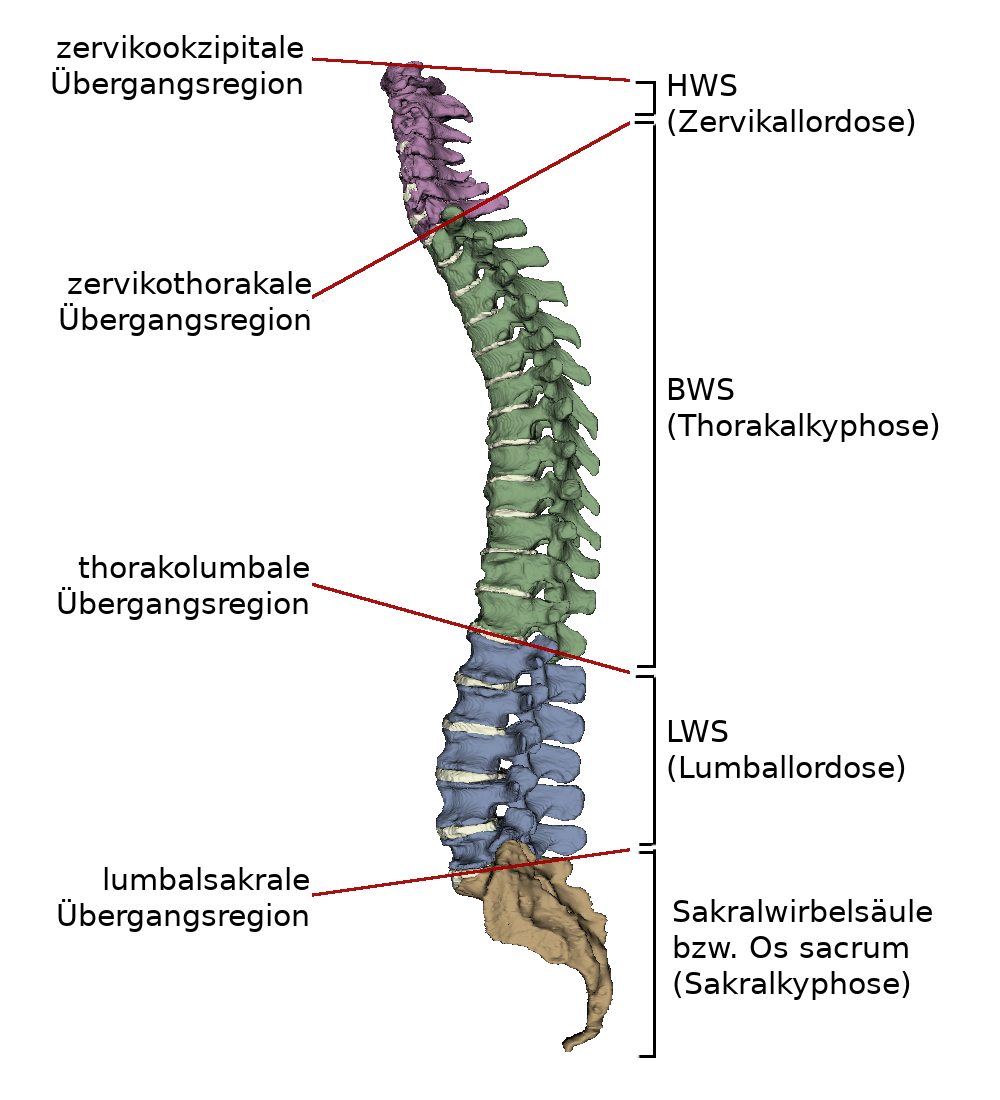
The latin term for the spinal column is Columna vertebralis. It runs from the head to the Pelvis, so from dorsal it represents the vertical axis of the human being. The spine of an adult human being is divided into four sections: Cervical spine (HWS), thoracic spine (BWS), lumbar spine (LWS) and sacral spine (Os sacrum). Accordingly, it also has four typical curvatures in the sagittal plane in humans. Each section is made up of individual vertebral bodies:
- 7 Cervical vertebrae (Vertebrae cervicales / C1-C7)
- 12 Thoracic vertebrae (Vertebrae thoraciace / T1-T12)
- 5 Lumbar vertebrae (Vertebrae lumbales / S1-S5)
- 5 Sacral vertebrae (Vertebrae sacrales / S1-S5 and Vertebrae coccygeae / Co1-Co5)
The different vertebrae each show a different structure of a vertebra. The vertebrae of the cervical, thoracic and lumbar spine are freely movable and connected to each other by the intervertebral discs. The sacral vertebrae, on the other hand, merge into the sacrum and coccyx in the course of life.
Shape of the spine
Seen from the side, the double "s" curvature of the spine is visible. The four sections thus each show a characteristic curvature:
- Cervical spine: cervical lordosis
- Thoracic spine: thoracic kyphosis
- Lumbar spine: lumbar lordosis
- Sacral spine: sacral kyphosis
These curvatures support the upright walk. Many people experience additional lateral curvatures due to strain. In the course of life, the spinal column becomes increasingly kyphotic. The spine has a spinal canal inside. This is formed by the structure of the vertebrae.
Deformations of the spine can cause wear and tear and muscle tension. In principle, there are three types of deformation: Scoliosis, kyphosis and lordosis. The kyphosis and lordosis are physiological to a certain degree.
Scoliosis
Here the natural curvature of the spine changes and begins to deform laterally. This twisting of the vertebral bodies around their own axis is called torsion. The cause of scoliosis cannot be found in 80% of cases. In some cases it can be traced back to:
- a disorder of the musculature (myopathic scoliosis)
- a partially congenital deformation of the vertebral bodies (osteopathic scoliosis)
- a disturbed nerve supply in the back (neuropathic scoliosis)
Kyphosis
In kyphosis, the upper part of the spine is curved forward. This deformation may be congenital or may only occur in the course of life, e.g. when the body tries to compensate for other deformities. The Cobb angle serves as a standard measure for assessing kyphosis. An example of a kyphotic form of a spine can be seen in the WebViewer.
Lordosis
A pathological change in the lordosis is described as a malposition of the lumbar spine, in which the abdominal area bulges forward due to the displacement of the pelvis. Late consequences would include herniated discs, spinal canal stenosis and gliding vertebrae. Causes can be shortened or untrained back muscles due to lack of exercise or incorrect posture. It inhibits the function of the abdominal muscles as antagonists by not relaxing.
Characteristic angles
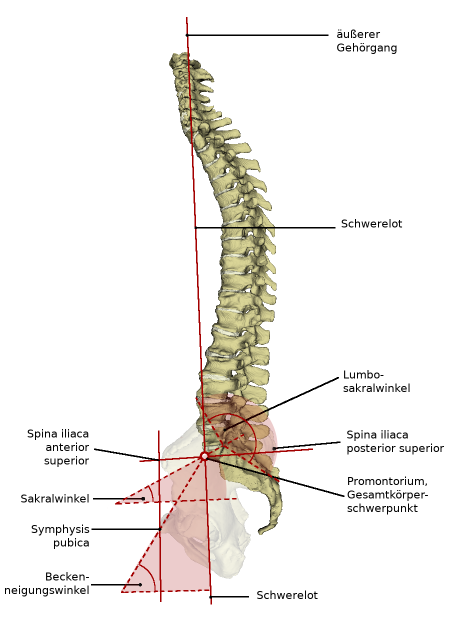
The installation of the spinal column in the pelvic girdle results in characteristic angles between imaginary axes. They help to uncover errors in the shape and position of the spinal column or trunk.
Sacral angle: this angle is approximately 30° and lies between the horizontal and the surface of the coccyx facing the head.
Lumbosacral angle: is approximately 135° and is located between the axes of the 5th lumbar vertebra and the 1st sacral vertebra. It decreases with a hollow back and increases with exaggerated pelvic straightening.
Angle of pelvic inclination (Inclinatio pelvis): approximately 60° with the body in an upright position. Angle between the virtual plane through the pelvis entrance and the horizontal. Since it increases or decreases when the pelvis is tilted forward or backward, the position of the pelvis can be determined relatively easily by palpable bone points.
Schwerelot: The gravity plumb line runs from the uppermost cervical vertebra to the upper end of the sacrum. Here, among other things, the external auditory canal, the spine of the 2nd cervical vertebra and the overall body centre of gravity is located directly ventral to the promontory.
Function
The spinal column stabilizes the trunk and makes it possible to maintain an upright posture. Furthermore, it is intended to create a large range of movement. Its characteristic curvature serves to dampen shocks, such as when walking or jumping. The anatomical structure of the spinal canal protects the spinal cord lying within it.
Movements of the spine
The movements of the spine are limited, but it does have a large range of motion. This is due to the fact that the individual vertebrae have a limited range of motion, but this is significantly increased by the stringing together of many vertebrae. This makes movements and deformations of the spinal column in all directions in space possible. A distinction is made between six movements:
- Flexion
- Extension
- Lateral flexion
- Lateral extension
- Rotation
These movements are influenced by the chest and back muscles, as well as by ligaments and joints.
Development
The spinal column is already formed in the fourth to tenth week of embryonic development. A newborn baby has a kyphotic spine. The lordotic curvatures of the cervical and lumbar spine are not yet present. This is due to the curved position of the fetus in the womb. Postnatally, the characteristic curvatures then develop over the course of life. First the cervical lordosis develops. This is due to the strengthening of the neck muscles. With learning to sit and stand, lumbar lordosis also develops. This develops further until the legs can be extended in the hip joints. The sacral vertebrae merge into the sacrum and coccyx in the course of development. This is completed by the age of 20, max. 25 years.
Diseases/Changes
Free exploration
- Spine
- Fall 1: Spina Bifida
- Fall 2: Kyphosis
Look at the structure of the spine in 3D and explore it freely. Afterwards you can test your acquired knowledge by the exercises.
In kyphosis, the upper part of the spine is curved forward. This deformation may be congenital or may develop in the course of life, for example, when the body tries to compensate for other deformities.