Schulterblatt/en: Unterschied zwischen den Versionen
(Übernehme Bearbeitung einer neuen Version der Quellseite) |
Becher (Diskussion | Beiträge) |
||
| (79 dazwischenliegende Versionen von 3 Benutzern werden nicht angezeigt) | |||
| Zeile 1: | Zeile 1: | ||
| − | + | The shoulder blade (lat. scapula) together with the [[Special:MyLanguage/Schlüsselbein|Clavicula]] forms the bony [[Special:MyLanguage/Schultergürtel|Shoulder Belt]]. The shoulder blade represents a flat three-sided bone, which occurs in pairs, just like the clavicle. It is located on the dorsal thorax and is completely embedded in muscle loops. It has a connection to the [[Special:MyLanguage/Oberarmknochen|Upper arm bone]] and to the clavicle. | |
| − | == | + | {{ArticleMenu_en|Links Übungsaufgaben=[[Special:MyLanguage/Übungsaufgaben: Obere Extremität|Upper extremity]][[Special:MyLanguage/Übungsaufgabe: Schultergürtel|Shoulder girdle]][[Special:MyLanguage/Übungsaufgabe: Schulterblatt|Shoulder blade]]| |
| + | Segmentereinbettung=<segmenter-embedding public wsemb-id="SchulterblattFrau" file="SchulterblattFrau.seg" height="300" width="400"/>| | ||
| + | Links Benachbarte Strukturen=[[Special:MyLanguage/Schlüsselbein|Clavicle]][[Special:MyLanguage/Oberarmknochen|Humerus]][[Special:MyLanguage/Rippen|Ribs]]| | ||
| + | Links Körperregionen=[[Special:MyLanguage/Schultergürtel|Shoulder girdle]][[Special:MyLanguage/Knochen Obere Extremität|Bones upper extremity]][[Special:MyLanguage/Obere Extremität|Upper extremity]]| | ||
| + | Links Organsystem=[[Special:MyLanguage/Platte Knochen|Plate bones]][[Special:MyLanguage/Knochen|Bones]][[Special:MyLanguage/Passiver Bewegungsapparat|Passive movement apparatus]][[Special:MyLanguage/Bewegungsapparat|Movement apparatus]]}} | ||
| − | <div class=" | + | ---- |
| − | + | ==Anatomy== | |
| + | <div class="thumb tright thumbinner"> | ||
| + | <div class="picture"> | ||
| + | <lightbox-embedding src="file:ScapulaVentralOhneBeschriftung.png" group="image-group-1" caption="View of the shoulder blade (Ventral)" width="400" height="300" style="width:300px; height:300px;float:left;margin:1px;background-color:#fff;border:1px solid #c8ccd1;display: flex;justify-content: center;"/> | ||
| + | </div> | ||
| + | <div class="gallery" style="margin-right:76px"> | ||
| + | <lightbox-embedding src="file:ScapulaDorsalOhneBeschriftung.png" group="image-group-1" caption="View of the shoulder blade (Dorsal)"/><lightbox-embedding src="file:ScapulaLateralOhneBeschriftung.png" group="image-group-1" caption="Ansicht der Scapula (Lateral)"/><lightbox-embedding src="file:PositionScapulaClavicula.png" group="image-group-1" caption="Position of clavicle to scapula"/> | ||
| + | </div> | ||
| + | <div class="thumbcaption"> | ||
| + | Views and position of the shoulder blade</div> | ||
</div> | </div> | ||
| − | |||
| − | |||
| − | |||
| − | |||
| − | |||
| − | |||
| − | + | The shoulder blade is a three-sided, flat bone. Together with the clavicle it forms the shoulder girdle. It has a variety of bony structures. The scapula is located dorsal to the thorax and is completely embedded in muscles. This musculature forms the connection to the [[Special:MyLanguage/Rumpf|Torso]]. On the shoulder blade, the [[Special:MyLanguage/Humeroscapular Gelenk|shoulder joint]] is formed at the connection to the humerus. Via the [[Special:MyLanguage/Acromioclavicular Gelenk|Acromioclavicular joint]] the shoulder blade and clavicle articulate with each other. | |
| − | + | In normal position, the scapula extends from the second to the seventh rib. The angulus inferior is located at the level of the spinous process of the seventh rib. The spina scapulae is located at the level of the spinous process of the third lumbar vertebra. In normal position, the scapula has a slight lateral rotation, so that the medial edge has an angle of about 3-5° to the spinous process. | |
| + | <div class="clear"></div> | ||
===Ränder=== | ===Ränder=== | ||
| + | <div class="thumb tright thumbinner"> | ||
| + | <div class="picture"> | ||
| + | <lightbox-embedding src="file:RänderSchulterblatt.png" group="image-group-3" caption="Characteristic edges of the shoulder blade" width="400" height="300" style="width:300px; height:300px;float:left;margin:1px;background-color:#fff;border:1px solid #c8ccd1;display: flex;justify-content: center;"/> | ||
| + | </div> | ||
| + | <div class="thumbcaption"> | ||
| + | Characteristic edges of the shoulder blade</div> | ||
| + | </div> | ||
| − | + | The flat bone is bordered by three edges. The upper edge is called margo superior. It represents the shortest edge of the bone and runs from the coracoid process (Proc. coracoideus) to the angulus superior. At this edge, medial to the base of the coracoid process, there is a distinct incision in the bony structure, the Incisura scapulae. | |
| − | + | The Margo medialis is the edge directed medially, towards the middle of the back. This is the longest side of the shoulder blade. It is located between the Angulus superior and the Angulus inferior. | |
| − | + | The outer margin, Margo lateralis, points laterally. This margin is the most massive of the three. It begins at the lower edge of the glenoid cavity and extends to the lower shoulder blade angle (Angulus inferior). | |
| + | <div class="clear"></div> | ||
| − | === | + | ===Angle=== |
| + | <div class="thumb tright thumbinner"> | ||
| + | <div class="picture"> | ||
| + | <lightbox-embedding src="file:WinkelSchulterblatt.png" group="image-group-3" caption="Characteristic angles of the shoulder blade" width="400" height="300" style="width:300px; height:300px;float:left;margin:1px;background-color:#fff;border:1px solid #c8ccd1;display: flex;justify-content: center;"/> | ||
| + | </div> | ||
| + | <div class="thumbcaption"> | ||
| + | Characteristic angles of the shoulder blade</div> | ||
| + | </div> | ||
| − | + | Because the shoulder blade resembles a triangle, three angles can be seen on it. | |
| − | + | Between the inner edge and the upper edge the angulus superior is enclosed. It represents the upper shoulder angle and is a rounded and flat bone area. | |
| − | + | Between the Margo medialis and the outer edge (Margo lateralis) is the lower angle of the shoulder blade, the Angulus inferior. The origin of the Musculus teres major and some fibres of the Musculus latissimus dorsi is located at the rear surface of this angle. | |
| − | + | The angulus lateralis is the lateral angle of the shoulder blade and is also called the "shoulder head" because it represents the most massive part of the scapula. It is located at the level of the cavitas glenoidalis. | |
| + | <div class="clear"></div> | ||
| − | === | + | ===Bony structure=== |
| + | <div class="thumb tright thumbinner"> | ||
| + | <div class="picture"> | ||
| + | <lightbox-embedding src="file:ScapulaVentral.png" group="image-group-2" caption="Anatomie der Scapula (Ventral)" width="400" height="300" style="width:300px; height:300px;float:left;margin:1px;background-color:#fff;border:1px solid #c8ccd1;display: flex;justify-content: center;"/> | ||
| + | </div> | ||
| + | <div class="gallery" style="margin-right:158px"> | ||
| + | <lightbox-embedding src="file:ScapulaDorsal.png" group="image-group-2" caption="Anatomy of the Shoulder blade (Dorsal)"/><lightbox-embedding src="file:ScapulaLateral.png" group="image-group-2" caption="Anatomy of the shoulder blade (Lateral)"/> | ||
| + | </div> | ||
| + | <div class="thumbcaption"> | ||
| + | Anatomy of the shoulder blade</div> | ||
| + | </div> | ||
| − | + | When viewed from ventral, the shoulder blade is characterized by a flat, slightly hollowed surface. This surface is the Facies posterior. The concave depression is called Fossa subscaolaris. Under certain circumstances Linea musculares may be visible on this surface. | |
| − | + | From dorsal view the back of the scapula, the Facies dorsalis (also Facies posterior) is visible. It is divided into two parts by the spine of scapula, the spina scapulae. Thus the upper smaller part, the Fossa supraspinata and the lower larger part, the Fossa infraspinata, are formed. The spina scapulae begins at the inner edge and extends to the lateral shoulder angle. It increases in height during its course and ends in the acromion (shoulder height or acromion). This is a flattened bone process. At the acromion there is an oval joint surface, the Facies articularis clavicularis, which forms the connection to the clavicle through the acromioclavicular joint. The angulus acromii is formed at the transition of the lateral edge of the acromion to the scapular spine. This is an easily palpable bone point. | |
| − | + | The glenoid cavity (cavitas glenoidales) is located in the angulus lateralis. There the scapula and humerus articulate with each other. At the upper edge of the glenoid cavity there is a small bony hump, this is the so-called tuberculum supraglenoidale. Below the cavitas glenoidales is the tuberculum infraglenoidale. In the direction of the large bone surface, the collum scapulae, the neck of the scapula, connects to the socket. Above the Cavitas glenoidales extends the coracoid process, the Processus Coracoideus, which originates from the Margo superior. It bends right-angled to lateroventral and ends flattened. The acromion and the raven-billed process together serve as a protection of the joint. | |
| − | + | The bony socket Cavitas glenoidalis of the [[Special:MyLanguage/Humeroscapular Gelenk|Humeroscapular joint]], a ball-and-socket joint, is considerably smaller than the humeral head. The socket is enlarged by a fibrocartilaginous joint lip, Labrum glenoidale. The surface of the Cavitas glenoidalis is about 6qcm. The weight of the upper extremity is about 4 kg. Since there are no stronger ligaments, the muscles that surround the joint must secure it. This is called a muscle-secured joint. The so-called "rotator cuff" is part of this muscular support and strengthens the joint capsule in particular. | |
| − | == | + | ==Movements== |
| − | + | The caput humeri is approximately spherical. The synovial joint capsule is attached to the labrum glenoidale of the scapula. | |
| − | + | Movements are possible in three degrees of freedom. These are called abduction and adduction, and are based on the resting position of the humeral caput in the scapula plane. The anteversion, the forward lifting of the arm, and its counter movement, the retroversion, are known. By a rotational component, a composite movement, the circumduction or circling, results with the involvement of the previously mentioned movements, whereby the arm practically describes a shell of a cone. | |
| − | + | In abduction movements, there is always a co-movement of the scapula; excessive co-movement of the scapula occurs in abduction over 90 degrees (elevation). | |
| − | == | + | ==Diseases== |
| − | *[[Special:MyLanguage/Scapulafraktur| | + | *[[Special:MyLanguage/Scapulafraktur|Fracture of the shoulder blade]] |
| − | *[[Special:MyLanguage/Rotatorenmanschettenruptur| | + | *[[Special:MyLanguage/Rotatorenmanschettenruptur|Rupture of the rotator cuff]] |
*[[Special:MyLanguage/Impingementsyndrom|Impingementsyndrom]] | *[[Special:MyLanguage/Impingementsyndrom|Impingementsyndrom]] | ||
| − | |||
| − | <div | + | ==Free exploration== |
| + | <div style="float:left;margin-right:1em;"><segmenter-embedding public wsemb-id="SchulterblattFrau" file="SchulterblattFrau.seg" height="300" width="400"/></div> | ||
| + | |||
| + | <div style="float:left;width:50%">Look at the structure of the shoulder blade in 3D and explore it freely. Afterwards you can test your acquired knowledge by the exercises.</div> | ||
| + | <div class="clear"></div> | ||
| + | |||
---- | ---- | ||
| − | + | <div class="clear aufgaben" style="margin-bottom:1em;"> | |
| + | <div class="menu_item"> | ||
| + | <div><li class="mw-ui-button button_new" >[[Special:MyLanguage/Übungsaufgaben|Exercises]]</li></div> | ||
| + | </div> | ||
| + | </div> | ||
| + | |||
| + | <div class="clear aufgaben"> | ||
| + | <div class="menu_item"> | ||
| + | <li class="button_article"><b>Further articles</b></li> | ||
| + | </div> | ||
| − | <div class =" | + | <div class="menu_item"> |
| + | <li class="mw-ui-button button_normal">[[Special:MyLanguage/Beckengürtel|Pelvic girdle]]</li> | ||
| + | </div> | ||
| + | |||
| + | <div class="menu_item"> | ||
| + | <li class="mw-ui-button button_normal">[[Special:MyLanguage/Humerus|Upper arm bone]]</li> | ||
| + | </div> | ||
| + | </div> | ||
| + | </div> | ||
| + | ---- | ||
| + | [[Category:Shoulder girdle]] | ||
| + | [[Category:Bones Upper Extremity]] | ||
| + | [[Category:Upper Extremity]] | ||
| + | [[Category:Body regions]] | ||
| + | |||
| + | </div> | ||
| + | <languages/> | ||
Aktuelle Version vom 16. Februar 2022, 09:15 Uhr
The shoulder blade (lat. scapula) together with the Clavicula forms the bony Shoulder Belt. The shoulder blade represents a flat three-sided bone, which occurs in pairs, just like the clavicle. It is located on the dorsal thorax and is completely embedded in muscle loops. It has a connection to the Upper arm bone and to the clavicle.
Inhaltsverzeichnis
Anatomy


The shoulder blade is a three-sided, flat bone. Together with the clavicle it forms the shoulder girdle. It has a variety of bony structures. The scapula is located dorsal to the thorax and is completely embedded in muscles. This musculature forms the connection to the Torso. On the shoulder blade, the shoulder joint is formed at the connection to the humerus. Via the Acromioclavicular joint the shoulder blade and clavicle articulate with each other.
In normal position, the scapula extends from the second to the seventh rib. The angulus inferior is located at the level of the spinous process of the seventh rib. The spina scapulae is located at the level of the spinous process of the third lumbar vertebra. In normal position, the scapula has a slight lateral rotation, so that the medial edge has an angle of about 3-5° to the spinous process.
Ränder

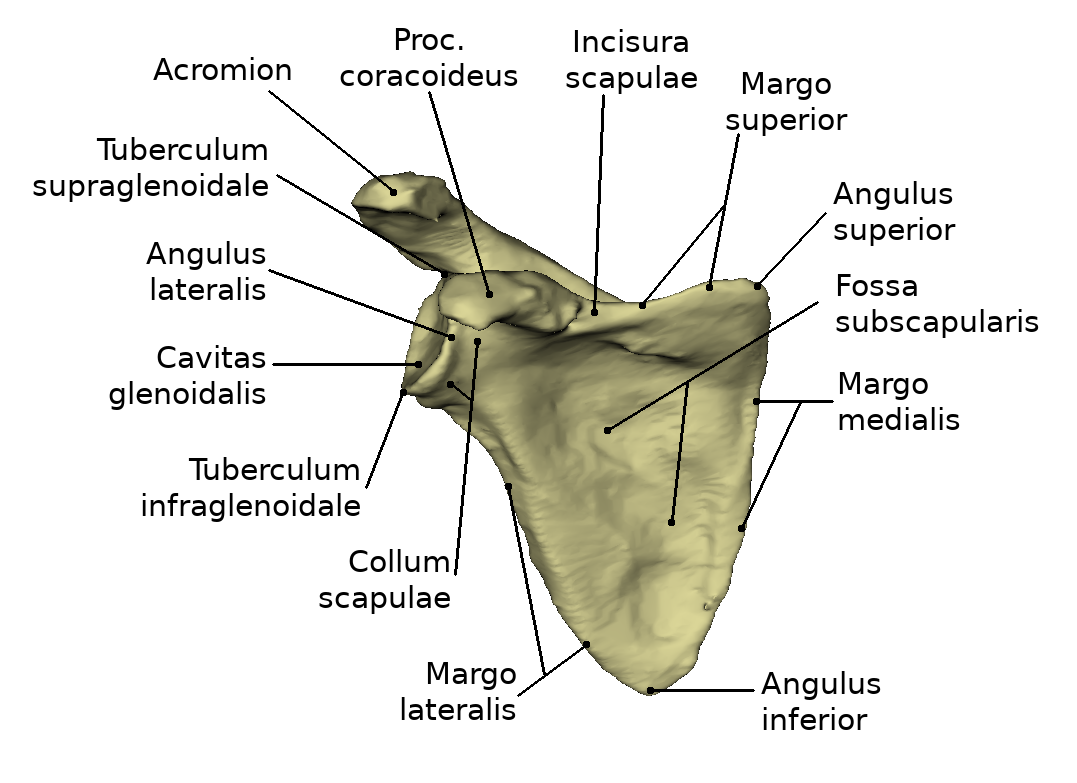
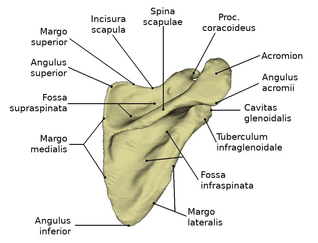
The flat bone is bordered by three edges. The upper edge is called margo superior. It represents the shortest edge of the bone and runs from the coracoid process (Proc. coracoideus) to the angulus superior. At this edge, medial to the base of the coracoid process, there is a distinct incision in the bony structure, the Incisura scapulae.
The Margo medialis is the edge directed medially, towards the middle of the back. This is the longest side of the shoulder blade. It is located between the Angulus superior and the Angulus inferior.
The outer margin, Margo lateralis, points laterally. This margin is the most massive of the three. It begins at the lower edge of the glenoid cavity and extends to the lower shoulder blade angle (Angulus inferior).
Angle
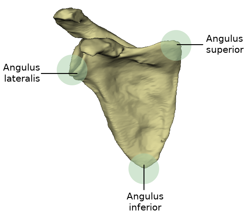
Because the shoulder blade resembles a triangle, three angles can be seen on it. Between the inner edge and the upper edge the angulus superior is enclosed. It represents the upper shoulder angle and is a rounded and flat bone area.
Between the Margo medialis and the outer edge (Margo lateralis) is the lower angle of the shoulder blade, the Angulus inferior. The origin of the Musculus teres major and some fibres of the Musculus latissimus dorsi is located at the rear surface of this angle.
The angulus lateralis is the lateral angle of the shoulder blade and is also called the "shoulder head" because it represents the most massive part of the scapula. It is located at the level of the cavitas glenoidalis.
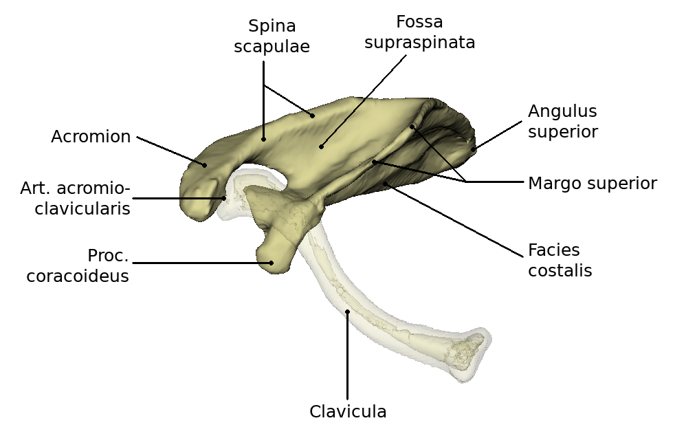
Bony structure

When viewed from ventral, the shoulder blade is characterized by a flat, slightly hollowed surface. This surface is the Facies posterior. The concave depression is called Fossa subscaolaris. Under certain circumstances Linea musculares may be visible on this surface.
From dorsal view the back of the scapula, the Facies dorsalis (also Facies posterior) is visible. It is divided into two parts by the spine of scapula, the spina scapulae. Thus the upper smaller part, the Fossa supraspinata and the lower larger part, the Fossa infraspinata, are formed. The spina scapulae begins at the inner edge and extends to the lateral shoulder angle. It increases in height during its course and ends in the acromion (shoulder height or acromion). This is a flattened bone process. At the acromion there is an oval joint surface, the Facies articularis clavicularis, which forms the connection to the clavicle through the acromioclavicular joint. The angulus acromii is formed at the transition of the lateral edge of the acromion to the scapular spine. This is an easily palpable bone point.
The glenoid cavity (cavitas glenoidales) is located in the angulus lateralis. There the scapula and humerus articulate with each other. At the upper edge of the glenoid cavity there is a small bony hump, this is the so-called tuberculum supraglenoidale. Below the cavitas glenoidales is the tuberculum infraglenoidale. In the direction of the large bone surface, the collum scapulae, the neck of the scapula, connects to the socket. Above the Cavitas glenoidales extends the coracoid process, the Processus Coracoideus, which originates from the Margo superior. It bends right-angled to lateroventral and ends flattened. The acromion and the raven-billed process together serve as a protection of the joint. The bony socket Cavitas glenoidalis of the Humeroscapular joint, a ball-and-socket joint, is considerably smaller than the humeral head. The socket is enlarged by a fibrocartilaginous joint lip, Labrum glenoidale. The surface of the Cavitas glenoidalis is about 6qcm. The weight of the upper extremity is about 4 kg. Since there are no stronger ligaments, the muscles that surround the joint must secure it. This is called a muscle-secured joint. The so-called "rotator cuff" is part of this muscular support and strengthens the joint capsule in particular.
Movements
The caput humeri is approximately spherical. The synovial joint capsule is attached to the labrum glenoidale of the scapula. Movements are possible in three degrees of freedom. These are called abduction and adduction, and are based on the resting position of the humeral caput in the scapula plane. The anteversion, the forward lifting of the arm, and its counter movement, the retroversion, are known. By a rotational component, a composite movement, the circumduction or circling, results with the involvement of the previously mentioned movements, whereby the arm practically describes a shell of a cone. In abduction movements, there is always a co-movement of the scapula; excessive co-movement of the scapula occurs in abduction over 90 degrees (elevation).