Zehenknochen/en: Unterschied zwischen den Versionen
Becher (Diskussion | Beiträge) (Die Seite wurde neu angelegt: „The big toe (hallux) consists of two limbs (phalanx proximalis and distalis), the remaining toes of three (phalanx proximalis, media and distalis). Like the me…“) |
Becher (Diskussion | Beiträge) |
||
| (39 dazwischenliegende Versionen von 3 Benutzern werden nicht angezeigt) | |||
| Zeile 1: | Zeile 1: | ||
The big toe (hallux) consists of two limbs (phalanx proximalis and distalis), the remaining toes of three (phalanx proximalis, media and distalis). Like the metatarsals, the proximal phalanges are divided into base, corpus and caput. | The big toe (hallux) consists of two limbs (phalanx proximalis and distalis), the remaining toes of three (phalanx proximalis, media and distalis). Like the metatarsals, the proximal phalanges are divided into base, corpus and caput. | ||
| − | + | ||
| − | + | {{ArticleMenu_en|Links Übungsaufgaben=[[Special:MyLanguage/Übungsaufgaben: Untere Extremität|Lower extremity]][[Special:MyLanguage/Übungsaufgabe: Zehenknochen|Toe bones]]| | |
| − | + | Segmentereinbettung=<segmenter-embedding public wsemb-id="AntetarsusFrau" file="AntetarsusFrau.seg" height="300" width="400"/>| | |
| − | + | Links Benachbarte Strukturen=[[Special:MyLanguage/Mittelfußknochen|Metatarsals]]| | |
| − | + | Links Körperregionen=[[Special:MyLanguage/Fußknochen|Foot bones]][[Special:MyLanguage/Knochen Untere Extremität|Bones lower extremity]][[Special:MyLanguage/Untere Extremität|Lower extremity]]| | |
| − | + | Links Organsystem=[[Special:MyLanguage/Röhrenknochen|Tubular bone]][[Special:MyLanguage/Knochen|Bones]][[Special:MyLanguage/Passiver Bewegungsapparat|Passive movement apparatus]][[Special:MyLanguage/Bewegungsapparat|Movement apparatus]]}} | |
| − | + | ||
| − | |||
| − | |||
| − | |||
| − | |||
| − | |||
| − | |||
---- | ---- | ||
| − | == | + | ==Anatomy== |
| − | + | {{ArticleGallery| | |
| − | + | Bild 1=<lightbox-embedding src="file:Antetarsus1.png" group="image-group-1" caption="View of the phalanges (Dorsum pedis)" width="400" height="300" style=""/>| | |
| − | <lightbox-embedding src="file:Antetarsus1.png" group="image-group-1" caption=" | + | Weitere Bilder=<lightbox-embedding src="file:Antetarsus2.png" group="image-group-1" caption="View of the phalanges (Planta pedis)"/> <lightbox-embedding src="file:AntetarsusKnochenBeschriftet1.png" group="image-group-1" caption="Anatomy of the phalanges (Dorsum pedis)"/> <lightbox-embedding src="file:AntetarsusKnochenBeschriftet2.png" group="image-group-1" caption="Anatomy of the phalanges (Planta pedis)"/> |
| − | + | |title=Anatomy of the phalanges}} | |
| − | |||
| − | <lightbox-embedding src="file:Antetarsus2.png" group="image-group-1" caption=" | ||
| − | |||
| − | |||
| − | |||
| − | |||
| − | + | {{ArticleGallery| | |
| − | + | Bild 1=<lightbox-embedding src="file:PhalanxBeschriftet.png" group="image-group-1" caption=Anatomy of the phalanges (Dorsum pedis)" width="400" height="300" style=""/>| | |
| − | <lightbox-embedding src="file:PhalanxBeschriftet.png" group="image-group-1" caption= | + | Weitere Bilder=<lightbox-embedding src="file:PhalanxBeschriftet2.png" group="image-group-1" caption="Anatomy of a toe bone (planta pedis)"/> <lightbox-embedding src="file:PhalanxProximalisBeschriftet1.png" group="image-group-1" caption="Anatomy of the proximal phalanx (dorsum pedis)"/> <lightbox-embedding src="file:PhalanxProximalisBeschriftet2.png" group="image-group-1" caption="Anatomy of the proximal phalanx (planta pedis)"/> <lightbox-embedding src="file:PhalanxMediaBeschriftet1.png" group="image-group-1" caption="Anatomy of the phalanx media (dorsum pedis)"/><lightbox-embedding src="file:PhalanxMediaBeschriftet2.png" group="image-group-1" caption="Anatomy of the phalanx media (planta pedis)"/> <lightbox-embedding src="file:PhalanxDistalisBeschriftet1.png" group="image-group-1" caption="Anatomy of the phalanx distalis (dorsum pedis)"/> <lightbox-embedding src="file:PhalanxDistalisBeschriftet2.png" group="image-group-1" caption="Anatomy of the phalanx distalis (planta pedis)"/> <lightbox-embedding src="file:OssaSesamoideaBeschriftet.png" group="image-group-1" caption="Anatomy of the sesamoid bone (Dorsum pedis)"/> |
| − | + | |title=Anatomy of a phalanges}} | |
| − | |||
| − | <lightbox-embedding src="file:PhalanxBeschriftet2.png" group="image-group-1" caption=" | ||
| − | |||
| − | |||
| − | <lightbox-embedding src="file:PhalanxMediaBeschriftet2.png" group="image-group-1" caption=" | ||
| − | |||
| − | |||
| − | |||
| − | |||
| − | + | The toe bones are formed by 14 tubular bones. These represent the phalanges of the foot. They are the end phalanges of the foot and therefore connect distally to the five metatarsal bones. The individual bony elements are divided between the individual toes. All toes consist of three phalanges, except the big toe, which consists of only two phalanges. | |
| − | + | Due to this division, the phalanges are called differently. The toe elements close to the body are the phalanx proximalis. The distal phalanges are called phalanx distalis. Between these are the phalanx media. In big toes the phalanx media is missing, so it consists only of the phalanx proximalis and phalanx distalis. | |
| − | + | The individual phalanges can also be structurally subdivided according to their affiliation to the tubular bones. A single one of these bone elements is subdivided into the head, the shaft and the base. | |
| − | + | The head is called caput phalangis and represents the distal end of the bone. It is flattened at the distal phalangis, where no other bone is adjacent. | |
| − | + | The proximal end of the tubular bone is called the basis phalangis. In the phalanx proximalis, this part has a concave base which is connected to the respective metatarsal bone. | |
| − | + | Between the base and the head of the phalanges is the shaft, the corpus phalangis. It is a somewhat narrower area and has a columnar-like shape. | |
| − | + | The proximal phalanx is also called the metatarsal phalanx and is the longest of the three bone links. Its base meets the respective metatarsal bone. The distal end articulates with the phalanx media at each toe. The exception to this is again the big toe. There the phalanx proximalis is directly connected to the phalanx distalis. | |
| − | + | The phalanx media is located in the middle of the phalanges series. It is medium in size and its base has a larger diameter than the distal end. At the head of this toe bone there are two small bony elevations. The proximal end connects to the phalanges. The distal end is followed by the phalanx distalis.<br> | |
| − | + | The distal limb of the entire lower extremity is the phalanx distalis. It is always the shortest of the phalanges. The base articulates with the phalanx media, except at the thumb where it is directly connected to the phalanx distalis. | |
| − | == | + | ==Free exploration== |
| − | <div style="float:left;margin-right:1em;"><segmenter-embedding | + | <div style="float:left;margin-right:1em;"><segmenter-embedding public wsemb-id="AntetarsusFrau" file="AntetarsusFrau.seg" height="300" width="400"/></div> |
| − | <div style="float:left;width:50%"> | + | <div style="float:left;width:50%">Look at the structure of the toe bones in 3D and explore them freely. Afterwards you can test your acquired knowledge by the exercises.</div> |
<div class="clear"></div> | <div class="clear"></div> | ||
---- | ---- | ||
| − | <div class=" | + | <div class="clear aufgaben" style="margin-bottom:1em;"> |
| − | < | + | <div class="menu_item"> |
| − | <div class=" | + | <li class="mw-ui-button button_new" >[[Special:MyLanguage/Übungsaufgaben|Exercises]]</li> |
| − | < | + | </div> |
| + | </div> | ||
| + | |||
| + | <div class="clear aufgaben"> | ||
| + | <div class="menu_item"> | ||
| + | <li class="button_article"><b>Further article</b></li> | ||
</div> | </div> | ||
| − | <div class=" | + | |
| − | + | <div class="menu_item"> | |
| + | <li class="mw-ui-button button_normal">[[Special:MyLanguage/Fingerknochen|Finger bones]]</li> | ||
</div> | </div> | ||
| + | <div class="menu_item"> | ||
| + | <li class="mw-ui-button button_normal">[[Special:MyLanguage/Wirbelsäule|Spine]]</li> | ||
| + | </div> | ||
| + | </div> | ||
| + | </div> | ||
<div class="clear"></div> | <div class="clear"></div> | ||
---- | ---- | ||
<languages/> | <languages/> | ||
| − | [[Category: | + | [[Category:Bones Lower Extremity]] |
| − | [[Category: | + | [[Category:Lower Extremity]] |
| − | [[Category: | + | [[Category:Body regions]] |
Aktuelle Version vom 17. Februar 2022, 14:55 Uhr
The big toe (hallux) consists of two limbs (phalanx proximalis and distalis), the remaining toes of three (phalanx proximalis, media and distalis). Like the metatarsals, the proximal phalanges are divided into base, corpus and caput.
Inhaltsverzeichnis
Anatomy


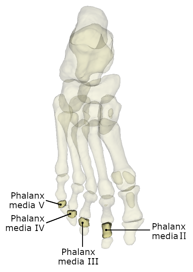


The toe bones are formed by 14 tubular bones. These represent the phalanges of the foot. They are the end phalanges of the foot and therefore connect distally to the five metatarsal bones. The individual bony elements are divided between the individual toes. All toes consist of three phalanges, except the big toe, which consists of only two phalanges. Due to this division, the phalanges are called differently. The toe elements close to the body are the phalanx proximalis. The distal phalanges are called phalanx distalis. Between these are the phalanx media. In big toes the phalanx media is missing, so it consists only of the phalanx proximalis and phalanx distalis.
The individual phalanges can also be structurally subdivided according to their affiliation to the tubular bones. A single one of these bone elements is subdivided into the head, the shaft and the base. The head is called caput phalangis and represents the distal end of the bone. It is flattened at the distal phalangis, where no other bone is adjacent.
The proximal end of the tubular bone is called the basis phalangis. In the phalanx proximalis, this part has a concave base which is connected to the respective metatarsal bone.
Between the base and the head of the phalanges is the shaft, the corpus phalangis. It is a somewhat narrower area and has a columnar-like shape.
The proximal phalanx is also called the metatarsal phalanx and is the longest of the three bone links. Its base meets the respective metatarsal bone. The distal end articulates with the phalanx media at each toe. The exception to this is again the big toe. There the phalanx proximalis is directly connected to the phalanx distalis.
The phalanx media is located in the middle of the phalanges series. It is medium in size and its base has a larger diameter than the distal end. At the head of this toe bone there are two small bony elevations. The proximal end connects to the phalanges. The distal end is followed by the phalanx distalis.
The distal limb of the entire lower extremity is the phalanx distalis. It is always the shortest of the phalanges. The base articulates with the phalanx media, except at the thumb where it is directly connected to the phalanx distalis.