Brustwirbelsäule/en: Unterschied zwischen den Versionen
Becher (Diskussion | Beiträge) (Die Seite wurde neu angelegt: „| <!--[segmenter_snapshot brustw 1]-->with annotations<!--[/]--> | <!--[segmenter_snapshot brustw 0]-->without annotations<!--[/]-->| <br> <br>“) |
Becher (Diskussion | Beiträge) |
||
| (49 dazwischenliegende Versionen von 3 Benutzern werden nicht angezeigt) | |||
| Zeile 1: | Zeile 1: | ||
| − | < | + | The spinal section between [[Special:MyLanguage/Halswirbelsäule|cervical spine]] and [[Special:MyLanguage/Lendenwirbelsäule|lumbar spine]] is the thoracic spine (BWS). It consists of twelve [[Special:MyLanguage/Aufbau Wirbel|structure of a vertebrae]]. These are stronger than the vertebrae in the cervical spine. The thoracic spine represents the longest part of the spinal column. Together with the ribs and sternum, the thoracic spine forms the bony thorax. |
| − | [[ | + | |
| + | {{ArticleMenu en|Links Übungsaufgaben=[[Special:MyLanguage/Übungsaufgaben: Rumpf|Torso]][[Special:MyLanguage/Übungsaufgabe: Wirbelsäule ventral|Spine]][[Special:MyLanguage/Übungsaufgabe: Brustwirbelsäule|Thoracic spine]]| | ||
| + | Segmentereinbettung=<segmenter-embedding public wsemb-id="BrustwirbelsaeuleMann" file="BrustwirbelsäuleMann.seg" width="400" height="300" />| | ||
| + | Links Benachbarte Strukturen=[[Special:MyLanguage/Halswirbelsäule|CERVICAL SPINE]][[Special:MyLanguage/Lendenwirbelsäule|LUMBAR SPINE]][[Special:MyLanguage/Rippen|Ribs]]| | ||
| + | Links Körperregionen=[[Special:MyLanguage/Wirbelsäule|Spine]][[Special:MyLanguage/Knochen Rumpf|Bones Torso]][[Special:MyLanguage/Rumpf|Torso]]| | ||
| + | Links Organsystem=[[Special:MyLanguage/Unregelmäßige Knochen|Irregular bones]][[Special:MyLanguage/Knochen|Bones]][[Special:MyLanguage/Passiver Bewegungsapparat|Passive musculoskeletal system]][[Special:MyLanguage/Bewegungsapparat|Musculoskeletal system]]}} | ||
| + | ---- | ||
| + | ==Anatomy== | ||
| + | {{ArticleGallery| | ||
| + | Bild 1=<lightbox-embedding src="file:BrustwirbelsäuleAnsicht1.png" group="image-group-1" caption="View of the thoracic spine (ventral)" width="400" height="300" style=""/>| | ||
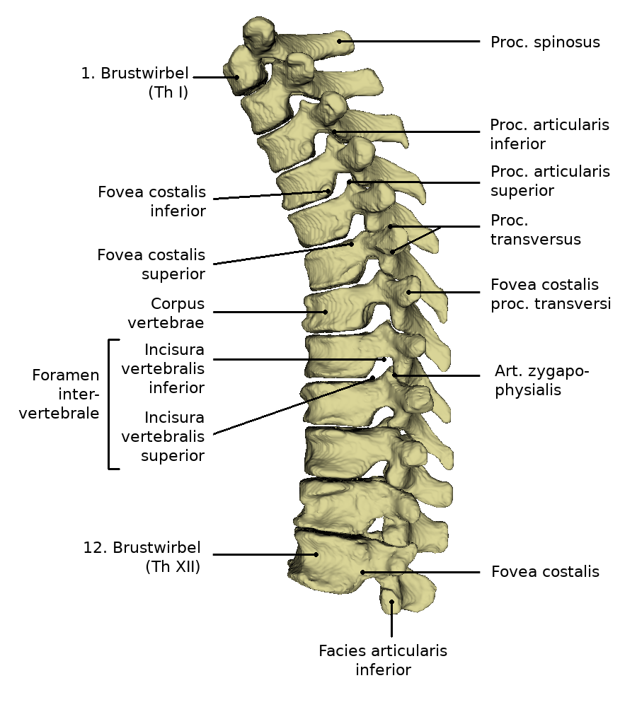
| + | Weitere Bilder=<lightbox-embedding src="file:BrustwirbelsäuleAnsicht2.png" group="image-group-1" caption="View of the thoracic spine (dorsal)"/><lightbox-embedding src="file:BrustwirbelsäuleAnsicht3.png" group="image-group-1" caption="View of the thoracic spine (left lateral)"/><lightbox-embedding src="file:Brustwirbelsäule beschriftet.png" group="image-group-1" caption="Anatomy of the thoracic spine"/> | ||
| + | |title=View and anatomy of the thoracic spine}} | ||
| + | |||
| + | |||
| + | {{ArticleGallery| | ||
| + | Bild 1=<lightbox-embedding src="file:BW2_1.png" group="image-group-1" caption="View of the second thoracic vertebra (ventral)" width="400" height="300" style=""/>| | ||
| + | Weitere Bilder=<lightbox-embedding src="file:BW2_2.png" group="image-group-1" caption="View of the second thoracic vertebra (cranial)"/><lightbox-embedding src="file:BW2_3.png" group="image-group-1" caption="View of the second thoracic vertebra (left lateral)"/><lightbox-embedding src="file:BW6_1.png" group="image-group-1" caption="View of the sixth thoracic vertebra (ventral)"/><lightbox-embedding src="file:BW6_2.png" group="image-group-1" caption="View of the sixth thoracic vertebra (cranial)"/><lightbox-embedding src="file:BW6_3.png" group="image-group-1" caption="View of the sixth thoracic vertebra (left lateral)"/><lightbox-embedding src="file:BW12_1.png" group="image-group-1" caption="View of the twelfth thoracic vertebra (ventral)"/><lightbox-embedding src="file:BW12_3.png" group="image-group-1" caption="View of the twelfth thoracic vertebra (cranial)"/><lightbox-embedding src="file:BW12_2.png" group="image-group-1" caption="View of the twelfth thoracic vertebra (lateral left)"/> | ||
| + | |title=View an anatomy of the thoracic spine}} | ||
| + | |||
| + | The thoracic vertebrae follow after the seventh cervical vertebra. Caudally of the twelfth thoracic vertebra the lumbar spine follows. In healthy people, the thoracic spine is bent backwards, which is known as physiological kyphosis.<br> | ||
| + | |||
| + | The thoracic vertebrae are stronger in comparison to the cervical vertebrae. They become stronger and more stable after caudally to bear the increasing body load. Seen laterally, the middle thoracic vertebrae have a lower height than the upper and lower thoracic vertebrae. The front side of the vertebrae is slightly higher than the back side. The surface facing the chest has a slightly hollowed out hollow. The spinous processes on the thoracic spine are long and triangular. They lie on top of each other like roof tiles, closing the gaps between the vertebral arches. Compared to the cervical and lumbar spine, the Foramen vertebrale is almost round and somewhat smaller. The transverse processes are bent away dorsally, thus creating space for the joint surfaces at the vertebral bodies and at the transverse processes.<br> | ||
| + | |||
| + | Between two vertebrae there is an intermediate hole. The spinal nerves run through this. At the level of the thoracic spine, these nerves innervate the rib cage, the corresponding muscles and the outer and inner skin of the chest wall. | ||
| + | |||
| + | The ribs are connected to the thoracic vertebrae. The uppermost joint pit for the first rib is completely supported by the first thoracic vertebra. Half of the joint cavity for the second rib is also located at this vertebra. At the upper and lower edges of the second to ninth thoracic vertebrae, there is a flat socket each, which together with the adjacent thoracic vertebra forms the articular surface for a rib head. The lower half of the articular cavity of the tenth rib is located on the upper side of the eleventh thoracic vertebra. The entire socket of the eleventh rib is also supported by the eleventh thoracic vertebra. The entire socket of the twelfth rib is also located on the twelfth thoracic vertebra. | ||
| + | |||
| + | ==Movement== | ||
| − | + | Through rib-vertebral joints it is possible to move the chest. This allows the rib cage to contract and expand again when breathing. The intercostal joints are formed by the vertebrae and the adjacent ribs. <br> | |
| − | + | The thoracic spine allows a lateral inclination of about 30° in the upper body. This movement is limited by the ribs, which meet at a certain point.<br> | |
| − | The | + | The rotation of the trunk around its own axis is also supported by the thoracic spine. Rotation is possible up to 33°. |
| − | + | ==Function== | |
| − | |||
| − | |||
| − | + | The trunk is stabilized by the thoracic spine. The individual ribs are held by the thoracic spine. Since the thoracic spine is involved in the construction of the thorax, the organs lying within are protected by the thoracic spine. In this part of the spine, too, the spinal cord is protected by the vertebrae. | |
| − | |||
| − | + | ==Free exploration== | |
| − | + | <div style="float:left;margin-right:1em;"><segmenter-embedding public wsemb-id="BrustwirbelsaeuleMann" src="segmenter:Nh2zaq9O804z" height="300px" width="500px"/></div> | |
| − | |||
| − | < | + | <div style="float:left;width:50%">Look at the structure of the thoracic spine in 3D and explore it freely. Afterwards you can test your acquired knowledge by the exercises.</div> |
| + | <div class="clear"></div> | ||
| − | |||
| − | |||
| − | |||
---- | ---- | ||
| − | + | {{ArticleMenuEnd en| | |
| − | + | Weiterer Artikel 1=[[Special:MyLanguage/Rippen|Ribs]]| | |
| − | + | Weiterer Artikel 2=[[Special:MyLanguage/Lendenwirbelsäule|Lumbar spine]]}} | |
| + | ---- | ||
| − | + | [[Category:Spine]] | |
| + | [[Category:Bones thorax]] | ||
| + | [[Category:Bones trunk]] | ||
| + | [[Category:Trunk]] | ||
| + | [[Category:Body regions]] | ||
| − | + | <languages/> | |
Aktuelle Version vom 11. Mai 2022, 07:27 Uhr
The spinal section between cervical spine and lumbar spine is the thoracic spine (BWS). It consists of twelve structure of a vertebrae. These are stronger than the vertebrae in the cervical spine. The thoracic spine represents the longest part of the spinal column. Together with the ribs and sternum, the thoracic spine forms the bony thorax.
Inhaltsverzeichnis
Anatomy


The thoracic vertebrae follow after the seventh cervical vertebra. Caudally of the twelfth thoracic vertebra the lumbar spine follows. In healthy people, the thoracic spine is bent backwards, which is known as physiological kyphosis.
The thoracic vertebrae are stronger in comparison to the cervical vertebrae. They become stronger and more stable after caudally to bear the increasing body load. Seen laterally, the middle thoracic vertebrae have a lower height than the upper and lower thoracic vertebrae. The front side of the vertebrae is slightly higher than the back side. The surface facing the chest has a slightly hollowed out hollow. The spinous processes on the thoracic spine are long and triangular. They lie on top of each other like roof tiles, closing the gaps between the vertebral arches. Compared to the cervical and lumbar spine, the Foramen vertebrale is almost round and somewhat smaller. The transverse processes are bent away dorsally, thus creating space for the joint surfaces at the vertebral bodies and at the transverse processes.
Between two vertebrae there is an intermediate hole. The spinal nerves run through this. At the level of the thoracic spine, these nerves innervate the rib cage, the corresponding muscles and the outer and inner skin of the chest wall.
The ribs are connected to the thoracic vertebrae. The uppermost joint pit for the first rib is completely supported by the first thoracic vertebra. Half of the joint cavity for the second rib is also located at this vertebra. At the upper and lower edges of the second to ninth thoracic vertebrae, there is a flat socket each, which together with the adjacent thoracic vertebra forms the articular surface for a rib head. The lower half of the articular cavity of the tenth rib is located on the upper side of the eleventh thoracic vertebra. The entire socket of the eleventh rib is also supported by the eleventh thoracic vertebra. The entire socket of the twelfth rib is also located on the twelfth thoracic vertebra.
Movement
Through rib-vertebral joints it is possible to move the chest. This allows the rib cage to contract and expand again when breathing. The intercostal joints are formed by the vertebrae and the adjacent ribs.
The thoracic spine allows a lateral inclination of about 30° in the upper body. This movement is limited by the ribs, which meet at a certain point.
The rotation of the trunk around its own axis is also supported by the thoracic spine. Rotation is possible up to 33°.
Function
The trunk is stabilized by the thoracic spine. The individual ribs are held by the thoracic spine. Since the thoracic spine is involved in the construction of the thorax, the organs lying within are protected by the thoracic spine. In this part of the spine, too, the spinal cord is protected by the vertebrae.