Aufbau Wirbel/en: Unterschied zwischen den Versionen
| Zeile 118: | Zeile 118: | ||
5. Ligament between the spinous processes | 5. Ligament between the spinous processes | ||
| − | + | 6. Exit canal of the nerve | |
| − | 6. | ||
</div> | </div> | ||
| + | <div class="clear"></div> --> | ||
==Freie Exploration== | ==Freie Exploration== | ||
Version vom 27. Mai 2020, 12:23 Uhr
The vertebrae are individual bony elements from which the Spine is constructed. In their outer shape, they follow a fundamentally identical form. Exceptions are the first two cervical vertebrae, Atlas and Axis, which have a different structure. The exact shape of the individual vertebrae depends on their position in the spinal column. Therefore, the individual vertebrae from Cervical spine, Thoracic spine and Lumbar spine differ slightly in their outer appearance.
Anatomy
Basic structure of a vertebra

The first two cervical vertebrae are the only vertebrae that deviate from the basic outer structure of the vertebrae. The bony structure of the remaining vertebrae is fundamentally similar. The vertebrae are composed of the following structures:
- a vertebral body (Corpus vertebrae)
- a vertebral arch (Arcus vertebrae)
- a vertebral foramen (Foramen vertebrale)
- a spinous process (Proc. spinosus)
- two transverse processes (Procc. Transversi or in the case of the lumbar vertebrae Procc. costales)
- four articular processes (Procc. Articulares)
Die Ausprägung der Wirbelstrukturen variiert in Anpassung an die Funktion und Belastung des jeweiligen Wirbelsäulenabschnittes.
Vertebral body


The vertebral bodies (Corpus vertebrae) have a round, almost oval shape and are based on a ring or hollow cylinder. They have a top and bottom cover plate. These areas are porous and form the connection to the adjacent Intervertebral discs.They are used for fastening straps. This means that in all vertebrae except the Atlas and the Axis, the vertebral body is a stably formed bone. The vertebral body is always aligned ventrally. In the thoracic and lumbar vertebrae, the vertebral body makes up the largest part of the vertebra and is the supporting part of the spine. The vertebral body is filled internally with red bone marrow and a tangled structure of fine bone balls, the sponglosa.
The first cervical vertebra (atlas) has no vertebral body. It is connected to the second cervical vertebra (axis) via a joint. Since the function of cervical vertebrae is primarily in mobility, the other cervical vertebrae have only a small vertebral body. The lumbar vertebrae, on the other hand, are subject to heavy strain and for this reason have a much larger and more massive vertebral body.
Vertebral arch
The vertebral arch feet (Pediculuc arcus vertebrae) and the arch plate (Lamina arcus vertebrae) together form the vertebral arch (Arcus vertebrae) at the back of the vertebra. In comparison to the vertebral body, it is significantly weaker. The horseshoe shape of the vertebral arch forms the vertebral hole (Foramen vertebralae), which is enclosed by the vertebral body and vertebral arch. The composition of the vertebrae results in the formation of the vertebral canal (Canalis vertebralis), which houses the spinal cord.
Spinous process
The spinous process (Proc. spinosus) protrudes dorsally along the cervical, thoracic and lumbar vertebrae. It is a bony extension that serves to attach ligaments and the back muscles. The individual muscles are supported by the joint leverage of the spinal and transverse processes. The only vertebra that does not have a spinous process is the atlas. Instead, it has a small bony hump (posterior tubercle). At the sacrum, the spinous process is no longer completely visible because it has receded. There is a bony crest with the name Crista sacralis mediana.
The spinous process varies slightly depending on its location in the spinal column. Thus it points slightly backwards and downwards at the second to sixth cervical vertebrae. There it is relatively short and forked in two prongs. The spinous process on the seventh cervical vertebra, on the other hand, juts out clearly and is a palpable bone point. The spinous process on the thoracic vertebrae is triangular and long. In the middle of the thoracic spine, the spinous processes are directed downwards, resulting in a tile-like arrangement. At the lumbar vertebrae there is a high spinous process which is horizontally directed backwards. These are laterally flattened.
If the spinous processes are missing in one or more vertebrae contrary to the norm, a vertebral gap is formed. This disease is called open spine or spina bifida.
Transverse processes
The transverse processes (procc. transversi or procc. costales in the lumbar vertebrae) are bony structures that protrude to both the left and right of the vertebral arch. Both ligaments and muscles are attached to them. In the area of the thoracic spine, they form the rib-vertebral joints. A hole is found in the transverse processes of the cervical vertebrae, the Foramen transversarium. Both blood vessels and nerves extend through this hole. At the lumbar vertebrae, the transverse processes (procc. costales) are rib rudiments and for this reason are not homologous to the other transverse processes at the cervical and thoracic spine (procc. transversi).
Articular processes
Each vertebra has four articular processes (Procc. Articulares). Two are directed upwards (superior) and two downwards (inferior). These bony structures form the true joints between the vertebrae. The inferior articular processes of the upper vertebra are always connected to the superior articular processes of the vertebra below. The thoracic and lumbar vertebrae have a wide structure, namely the teat process (procc. mamilaris). This is positioned on the upward pointing articular processes.
Typical vertebrae from different areas of the spine
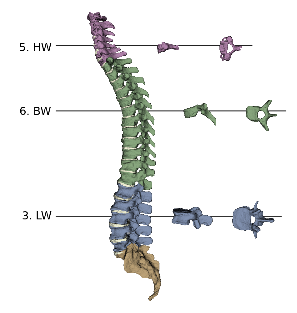
The vertebrae vary in the different regions of the spine in terms of their size and their special characteristics. On the one side, the vertebral bodies increase in size from cranial to caudal and on the other, the vertebral foramen gradually become smaller as the spinal cord becomes narrower. Furthermore, the appearance of the vertebral arches and the adjacent processes changes.
The figure on the right shows:
- A - Fifth cervical vertebrae
- B - Sixth thoracic vertebrae
- C - Third lumbar vertebrae
motion segment
Spinal motion segment is the functional unit that two adjacent vertebrae including the intermediate spinal disc (synchondrosis) and the adjacent intersegmental musculature together form.
Furthermore, it describes the comprehensive interaction of all bony, cartilaginous, ligamentary, and muscular components of a defined spinal segment.
Thus, the spinal motion segment forms the smallest functional unit of the spinal column.