Oberarmknochen/en: Unterschied zwischen den Versionen
Becher (Diskussion | Beiträge) (Die Seite wurde neu angelegt: „The <i>Caput humeri</i> (1) is approximately spherical. The synovial joint capsule is attached to the <i>Labrum glenoidale</i> of the scapula. It extends like…“) |
Becher (Diskussion | Beiträge) |
||
| (59 dazwischenliegende Versionen von 3 Benutzern werden nicht angezeigt) | |||
| Zeile 1: | Zeile 1: | ||
| − | < | + | The upper arm bone (lat. Humerus) is a strong, straight and long tubular bone. It is the largest bone in the human arm. It forms the connection between shoulder and elbow. In general, the humerus is divided into the proximal end piece, the humeral shaft and the distal end piece. |
| + | |||
| + | <div class="button_style"> | ||
| + | <div class ="center"> | ||
| + | <div class="dropdown"> | ||
| + | <div class="floatright" style="margin:0.4em;">[[File:PfeilDropdown.png|20px|link=|Exercises]]</div> | ||
| + | <div class="dropbtnart">[[Special:MyLanguage/Übungsaufgaben|Exercises]]</div> | ||
| + | <div class="dropdown-content"> | ||
| + | <div>[[Special:MyLanguage/Übungsaufgaben: Obere Extremität|Upper Extremity]]</div> | ||
| + | <div>[[Special:MyLanguage/Übungsaufgabe: Oberarmknochen|Humerus]]</div> | ||
| + | </div> | ||
| + | </div> | ||
| + | </div> | ||
| + | </div> | ||
| + | |||
| + | <div class="box"> | ||
| + | <div class="pic">__TOC__</div> | ||
| + | <div class="segmenter"><segmenter-embedding public wsemb-id="HumerusFrau" file="HumerusFrau.seg" height="300" width="400"/></div> | ||
| + | </div> | ||
| + | |||
| + | <div class="dropdown"> | ||
| + | <div class="floatright" style="margin:0.4em;">[[File:PfeilDropdown.png|20px|link=|Neighbouring structures]]</div> | ||
| + | <div class="dropbtnart">Neighbouring structures</div> | ||
| + | <div class="dropdown-content"> | ||
| + | <div>[[Special:MyLanguage/Schultergürtel|Shoulder girdle]]</div> | ||
| + | <div>[[Special:MyLanguage/Unterarmknochen|Forearm bones]]</div> | ||
| + | </div> | ||
| + | </div> | ||
| + | |||
| + | <div class="dropdown"> | ||
| + | <div class="floatright" style="margin:0.4em;">[[File:PfeilDropdown.png|20px|link=|Body regions]]</div> | ||
| + | <div class="dropbtnart">[[Special:MyLanguage/Körperregionen|Body regions]]</div> | ||
| + | <div class="dropdown-content"> | ||
| + | <div>[[Special:MyLanguage/Schultergürtel|Shoulder girdle]]</div> | ||
| + | <div>[[Special:MyLanguage/Knochen Obere Extremität|Bones Upper Extremity]]</div> | ||
| + | <div>[[Special:MyLanguage/Obere Extremität|Upper Extremity]]</div> | ||
| + | </div> | ||
| + | </div> | ||
| + | |||
| + | <div class="dropdown"> | ||
| + | <div class="floatright" style="margin:0.4em;">[[File:PfeilDropdown.png|20px|link=|Organ system]]</div> | ||
| + | <div class="dropbtnart">[[Special:MyLanguage/Organsystem|Organ system]]</div> | ||
| + | <div class="dropdown-content"> | ||
| + | <div>[[Special:MyLanguage/Röhrenknochen|Tubular bones]]</div> | ||
| + | <div>[[Special:MyLanguage/Knochen|Bones]]</div> | ||
| + | <div>[[Special:MyLanguage/Passiver Bewegungsapparat|Passive movement apperatus]]</div> | ||
| + | <div>[[Special:MyLanguage/Bewegungsapparat|Movement apperatus]]</div> | ||
| + | </div> | ||
| + | </div> | ||
| + | ---- | ||
| + | ==Anatomy== | ||
| + | |||
| + | {{ArticleGallery| | ||
| + | Bild 1=<lightbox-embedding src="file:Humerus.png" group="image-group-1" caption="Upper arm bone" width="400" height="300" style=""/>| | ||
| + | Weitere Bilder=<lightbox-embedding src="file:OberarmknochenDorsal.png" group="image-group-1" caption="View of the humerus (dorsal)"/> | ||
| + | <lightbox-embedding src="file:Humeruskopf.png" group="image-group-1" caption="View of the humeral head"/> | ||
| + | <lightbox-embedding src="file:OberarmknochenKaudal.png" group="image-group-1" caption="View of the distal joint surface of the humerus"/> | ||
| + | |title=Views of the right humerus}} | ||
| + | |||
| + | {{ArticleGallery| | ||
| + | Bild 1=<lightbox-embedding src="file:HumerusLateral.png" group="image-group-1" caption="Upper arm bone" width="400" height="300" style=""/>| | ||
| + | Weitere Bilder=<lightbox-embedding src="file:HumerusMedial.png" group="image-group-1" caption="Anatomy of the humerus (medial)"/> <lightbox-embedding src="file:HumerusDorsal.png" group="image-group-1" caption="Humerus dorsal"/> <lightbox-embedding src="file:HumerusVentral.png" group="image-group-1" caption="Anatomy of the humerus (ventral)"/><lightbox-embedding src="file:HumeruskopfKranial.png" group="image-group-1" caption="Anatomy of the humeral head (kranial)"/><lightbox-embedding src="file:DistalerHumerusKaudal.png" group="image-group-1" caption="Anatomy of the distal joint surface of the humerus"/> | ||
| + | |title=Anatomy of the right humerus}} | ||
| − | |||
| − | The humerus is articulately connected to [[Special:MyLanguage/Schulterblatt| | + | The humerus is articulately connected to [[Special:MyLanguage/Schulterblatt|shoulder blade]], [[Special:MyLanguage/Elle|ulna]] and [[Special:MyLanguage/Speiche|radius]]. |
| − | + | It consists of the corpus and a proximal and distal extremity. The proximal end forms the caput humeri with the adjacent neck, collum humeri. It appears slightly retracted and is also called Collum anatomicum. The distal end is formed by the condyle humeri.<br> | |
| + | At the anterior surface of the proximal extremity, the tubercle majus is located laterally and the tubercle minus medially. Between the two, there is a strong depression, the sulcus intertubercularis, which serves as a sliding rail for the tendon of the long caput of the biceps brachii.<br> | ||
| + | At the posterior surface of the corpus humeri lies the sulcus n. radialis. At the distal extremity there is the medially more powerful bony protrusion Epicondylus medialis, laterally the weaker Epicondylus lateralis.<br> | ||
| + | Trochlea humeri and Capitulum humeri form the Condylus humeri and serve as the joint connection with the forearm bones. Medially from the trochlea humeri runs a shallow channel, the sulcus n. ulnaris. On the posterior surface above the trochlea there is a deep pit, the olecranon fossa. | ||
| + | The humerus is torqued at its proximal end, i.e. the caput is twisted about 20° backwards in relation to the axis through the epicondyli humeri (torsion). Between the longitudinal axis of the humerus and the caput there is an angle of about 130° on average, while at the distal end, between the transverse joint axis and the longitudinal axis of the humeral shaft, there is an angle of 76 to 89°.<br> | ||
| + | The bony socket (Cavitas glenoidalis) of the Articulatio humeri, a ball-and-socket joint, is considerably smaller than the humeral head. The socket is enlarged by a fibrocartilaginous joint lip, labrum glenoidale. The surface area of the cavitas glenoidalis is approximately 6 cm². The weight of the upper extremity is approximately 4 kg. Since there are no stronger ligaments, the muscles that surround the joint must secure it. This is therefore called a muscle-secured joint. The so-called "rotator cuff" is part of this muscular support and strengthens the joint capsule in particular. | ||
| + | The caput humeri is approximately spherical. The synovial joint capsule is attached to the labrum glenoidale of the scapula. The synovial joint capsule is attached to the labrum glenoidale of the scapula and extends along the intracapsular tendon of the long head of the biceps like a sack, enclosing it with a vagina synovialis intertubercularis, which is located in the sulcus intertubercularis. | ||
| − | + | ==Function== | |
| − | + | The humerus connects the shoulder and forearm. It is involved in the formation of the shoulder joint and the elbow joint. The humerus serves as the base and origin of many muscles that are needed to move the arm and shoulder. | |
| − | + | ==Movement== | |
| + | Movements in three degrees of freedom are possible. One speaks of abduction and adduction, whereby one assumes the resting position of the Caput humeri in the scapula plane. One knows the anteversion, the forward lifting of the arm and its counter movement, the retroversion. By a rotational component, a composite movement, the circumduction or circling, results with the involvement of the previously mentioned movements, whereby the arm practically describes a conical shell. | ||
| − | + | Abduction movements always result in a co-motion of the scapula; excessive co-motion of the scapula occurs with an abduction of more than 90 degrees (elevation). | |
| − | + | ==Diseases== | |
| + | * [[Special:MyLanguage/Humerusfraktur|frakture of the humerus]] | ||
| − | + | ==Free exploration== | |
| + | <div style="float:left;margin-right:1em;"><segmenter-embedding public wsemb-id="HumerusFrau" file="HumerusFrau.seg" height="300" width="400"/></div> | ||
| − | + | <div style="float:left;width:50%">Look at the structure of the upper arm bone in 3D and explore it freely. Afterwards you can test your acquired knowledge by the exercises.</div> | |
| + | <div class="clear"></div> | ||
| − | + | ---- | |
| + | <div class="clear aufgaben" style="margin-bottom:1em;"> | ||
| + | <div class="menu_item"> | ||
| + | <li class="mw-ui-button button_new" >[[Special:MyLanguage/Übungsaufgaben|Exercises]]</li> | ||
| + | </div> | ||
| + | </div> | ||
| − | + | <div class="clear aufgaben"> | |
| + | <div class="menu_item"> | ||
| + | <li class="mw-ui-button button_normal"><b>Further article</b></li> | ||
| + | </div> | ||
| + | <div class="menu_item"> | ||
| + | <li class="mw-ui-button button_normal">[[Special:MyLanguage/Oberschenkelknochen|Femur]]</li> | ||
| + | </div> | ||
| − | -- | + | <div class="menu_item"> |
| + | <li class="mw-ui-button button_normal">[[Special:MyLanguage/Handknochen|Hand bones]]</li> | ||
| + | </div> | ||
| + | </div> | ||
| − | + | <div class="clear"></div> | |
| + | ---- | ||
| − | [[ | + | [[Category:Bones Upper Extremity]] |
| + | [[Category:Upper Extremity]] | ||
| + | [[Category:Body regions]] | ||
| − | + | </div> | |
| + | <languages/> | ||
Aktuelle Version vom 5. Januar 2022, 13:58 Uhr
The upper arm bone (lat. Humerus) is a strong, straight and long tubular bone. It is the largest bone in the human arm. It forms the connection between shoulder and elbow. In general, the humerus is divided into the proximal end piece, the humeral shaft and the distal end piece.
Inhaltsverzeichnis
Anatomy


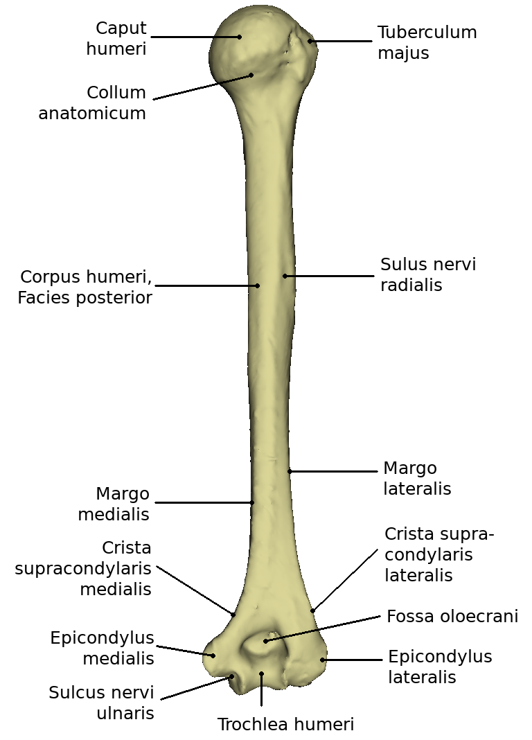


The humerus is articulately connected to shoulder blade, ulna and radius.
It consists of the corpus and a proximal and distal extremity. The proximal end forms the caput humeri with the adjacent neck, collum humeri. It appears slightly retracted and is also called Collum anatomicum. The distal end is formed by the condyle humeri.
At the anterior surface of the proximal extremity, the tubercle majus is located laterally and the tubercle minus medially. Between the two, there is a strong depression, the sulcus intertubercularis, which serves as a sliding rail for the tendon of the long caput of the biceps brachii.
At the posterior surface of the corpus humeri lies the sulcus n. radialis. At the distal extremity there is the medially more powerful bony protrusion Epicondylus medialis, laterally the weaker Epicondylus lateralis.
Trochlea humeri and Capitulum humeri form the Condylus humeri and serve as the joint connection with the forearm bones. Medially from the trochlea humeri runs a shallow channel, the sulcus n. ulnaris. On the posterior surface above the trochlea there is a deep pit, the olecranon fossa.
The humerus is torqued at its proximal end, i.e. the caput is twisted about 20° backwards in relation to the axis through the epicondyli humeri (torsion). Between the longitudinal axis of the humerus and the caput there is an angle of about 130° on average, while at the distal end, between the transverse joint axis and the longitudinal axis of the humeral shaft, there is an angle of 76 to 89°.
The bony socket (Cavitas glenoidalis) of the Articulatio humeri, a ball-and-socket joint, is considerably smaller than the humeral head. The socket is enlarged by a fibrocartilaginous joint lip, labrum glenoidale. The surface area of the cavitas glenoidalis is approximately 6 cm². The weight of the upper extremity is approximately 4 kg. Since there are no stronger ligaments, the muscles that surround the joint must secure it. This is therefore called a muscle-secured joint. The so-called "rotator cuff" is part of this muscular support and strengthens the joint capsule in particular.
The caput humeri is approximately spherical. The synovial joint capsule is attached to the labrum glenoidale of the scapula. The synovial joint capsule is attached to the labrum glenoidale of the scapula and extends along the intracapsular tendon of the long head of the biceps like a sack, enclosing it with a vagina synovialis intertubercularis, which is located in the sulcus intertubercularis.
Function
The humerus connects the shoulder and forearm. It is involved in the formation of the shoulder joint and the elbow joint. The humerus serves as the base and origin of many muscles that are needed to move the arm and shoulder.
Movement
Movements in three degrees of freedom are possible. One speaks of abduction and adduction, whereby one assumes the resting position of the Caput humeri in the scapula plane. One knows the anteversion, the forward lifting of the arm and its counter movement, the retroversion. By a rotational component, a composite movement, the circumduction or circling, results with the involvement of the previously mentioned movements, whereby the arm practically describes a conical shell.
Abduction movements always result in a co-motion of the scapula; excessive co-motion of the scapula occurs with an abduction of more than 90 degrees (elevation).