Speiseröhre/en: Unterschied zwischen den Versionen
Becher (Diskussion | Beiträge) (Die Seite wurde neu angelegt: „The human esophagus is approximately 23-27 centimeters long and has a diameter of 1-2 centimeters. It can be divided into three sections:“) |
Becher (Diskussion | Beiträge) |
||
| (26 dazwischenliegende Versionen von 2 Benutzern werden nicht angezeigt) | |||
| Zeile 1: | Zeile 1: | ||
| − | + | The '''esophagus''' (Latin '''Oesophagus''') is a muscular tube that, as part of the digestive system, carries swallowed food from the pharynx to the [[Special:MyLanguage/Magen|stomach]] by means of peristaltic movements. | |
| − | The ''' | ||
<div class="button_style"> | <div class="button_style"> | ||
| Zeile 65: | Zeile 64: | ||
The human esophagus is approximately 23-27 centimeters long and has a diameter of 1-2 centimeters. It can be divided into three sections: | The human esophagus is approximately 23-27 centimeters long and has a diameter of 1-2 centimeters. It can be divided into three sections: | ||
| − | + | The '''pars cervicalis''' extends from HWK 6 to BWK 1 and lies anterior to the spine in the region of the neck. <br> | |
| − | + | The pars thoracica is the longest section and is located in the upper and posterior mediastinum (mediastinum superius and mediastinum posterius). It runs from BWK 1 to about BWK 11, where the passage through the diaphragm is located. <br> | |
| − | + | The shortest section is the '''pars abdominalis'''. This extends from the piercing of the diaphragm to the entrance of the stomach (lat. Cardia). In addition, it is already in the peritoneal cavity (lat. Cavitas peritonealis). | |
| − | === | + | ===Projection onto the trunk=== |
| − | + | Due to its close relationship with neighboring organs, the esophagus has three narrowing points. | |
| − | + | The '''esophageal orifice''' refers to the uppermost and narrowest part (''constrictio pharyngooesophagealis''). This is located in the pars cervicalis at the level of the cricoid cartilage of the larynx and measures approximately 14 millimeters in maximum diameter. Starting from the cricoid cartilage of the larynx, muscles surround the esophageal orifice and close it in the form of a sphincter, which is closed at rest. | |
| − | + | The middle constriction (''Constrictio partis thoracicae'') in the pars thoracica is also called '''aortic stenosis'''. This is caused by the constriction of the esophagus by the left main bronchus and the aortic arch in the area of BWK 4/5. The maximum diameter here is 14 millimeters. | |
| − | + | The inferior constriction (''constrictio phrenica'') results from the esophagus passing through the diaphragm at the beginning of the pars abdominalis and is called '''diaphragmatic constriction'''. Functional closure of the esophagus is ensured by the adjacent longitudinal tension of the musculature and by the veins of the esophageal wall. At rest (outside the act of swallowing), the lower part of the esophagus (pars abdominalis) is permanently closed. | |
| − | === | + | ===Position in horizontal section=== |
| − | In | + | In the ventral view of the [[Special:MyLanguage/Thorax|thorax]], the esophagus lies predominantly in the lower part and is slightly displaced to the right with respect to the median line by the aorta running to its left. A little below the ''Proc. xiphoideus sterni'' it enters the peritoneal cavity through the [[Special:MyLanguage/Zwerchfell|diaphragm]]. |
| − | === | + | ===Relationship to neighbouring organs=== |
| − | + | Immediately on the anterior side of the esophagus runs the [[Special:MyLanguage/Luftröhre|trachea]] until it divides into the two main bronchi (Latin ''bifurcatio tracheae''). The anterior wall of the esophagus is connected to the posterior wall of the trachea by a multitude of connective tissue reins. <br> | |
| − | + | Below the structure of the tracheal bifurcation, in the lower mediastinum (Latin ''mediastinum inferius''), the esophagus lies close to the left atrium and the thoracic aorta. Furthermore, the aorta initially runs caudally to the left of the esophagus and comes to rest behind the esophagus shortly before it enters the diaphragm. | |
| − | === | + | ===Position In situ=== |
| − | + | The esophagus has characteristic curvatures in addition to strictures. | |
| − | + | The '''upper curvature''' runs in the view from ventral in the pars cervicalis to the left. The '''middle curvature''' is in the pars thoracica and runs to the right due to the nearby thoracic aorta. The '''inferior curvature''' in the pars abdominalis now turns back to the left. | |
| − | + | If the esophagus is viewed in the sagittal plane, it can be seen that it follows the course of the spine and is curved forward in a slightly concave manner. | |
| − | == | + | ==Diseases== |
| − | *[[Special:MyLanguage/Divertikel| | + | *[[Special:MyLanguage/Divertikel|Diverticulum]] |
| − | *[[Special:MyLanguage/Ösophagitis| | + | *[[Special:MyLanguage/Ösophagitis|Esophagitis]] |
| − | *[[Special:MyLanguage/gastroösophageale Refluxkrankheit| | + | *[[Special:MyLanguage/gastroösophageale Refluxkrankheit|gastroesophageal reflux disease]] |
| − | *[[Special:MyLanguage/Speiseröhrenkrebs| | + | *[[Special:MyLanguage/Speiseröhrenkrebs|Esophageal cancer]] |
| − | == | + | ==Free exploration== |
| − | <div style="float:left;margin-right:1em;"><segmenter-embedding wsemb-id="Speiseroehre" file="Speiseroehre.seg" height="300" width="400"/></div> | + | <div style="float:left;margin-right:1em;"><segmenter-embedding public wsemb-id="Speiseroehre" file="Speiseroehre.seg" height="300" width="400"/></div> |
| − | <div style="float:left;width:50%"> | + | <div style="float:left;width:50%">Look at the structure of the esophagus in 3D and explore it freely. Afterwards, you can check your learned knowledge through the exercise tasks.</div> |
<div class="clear"></div> | <div class="clear"></div> | ||
| Zeile 121: | Zeile 120: | ||
<div class="clear aufgaben" style="margin-bottom:1em;"> | <div class="clear aufgaben" style="margin-bottom:1em;"> | ||
<div class="menu_item"> | <div class="menu_item"> | ||
| − | <div | + | <div><li class="mw-ui-button button_new" >[[Special:MyLanguage/Übungsaufgaben|Exercises]]</li></div> |
</div> | </div> | ||
</div> | </div> | ||
| Zeile 127: | Zeile 126: | ||
<div class="clear aufgaben"> | <div class="clear aufgaben"> | ||
<div class="menu_item"> | <div class="menu_item"> | ||
| − | <li class="button_article button_normal"><b> | + | <li class="button_article button_normal"><b>Further article</b></li> |
</div> | </div> | ||
<div class="menu_item"> | <div class="menu_item"> | ||
| − | <li class="mw-ui-button button_normal">[[Special:MyLanguage/ | + | <li class="mw-ui-button button_normal">[[Special:MyLanguage/Magen|Stomach]]</li> |
</div> | </div> | ||
<div class="menu_item"> | <div class="menu_item"> | ||
| − | <li class="mw-ui-button button_normal">[[Special:MyLanguage/Kehlkopf| | + | <li class="mw-ui-button button_normal">[[Special:MyLanguage/Kehlkopf|Larynx]]</li> |
</div> | </div> | ||
</div> | </div> | ||
| Zeile 141: | Zeile 140: | ||
---- | ---- | ||
| − | [[Category: | + | [[Category:Digestive system]] |
| + | <languages/> | ||
Aktuelle Version vom 15. September 2022, 07:20 Uhr
The esophagus (Latin Oesophagus) is a muscular tube that, as part of the digestive system, carries swallowed food from the pharynx to the stomach by means of peristaltic movements.
Anatomy

Division
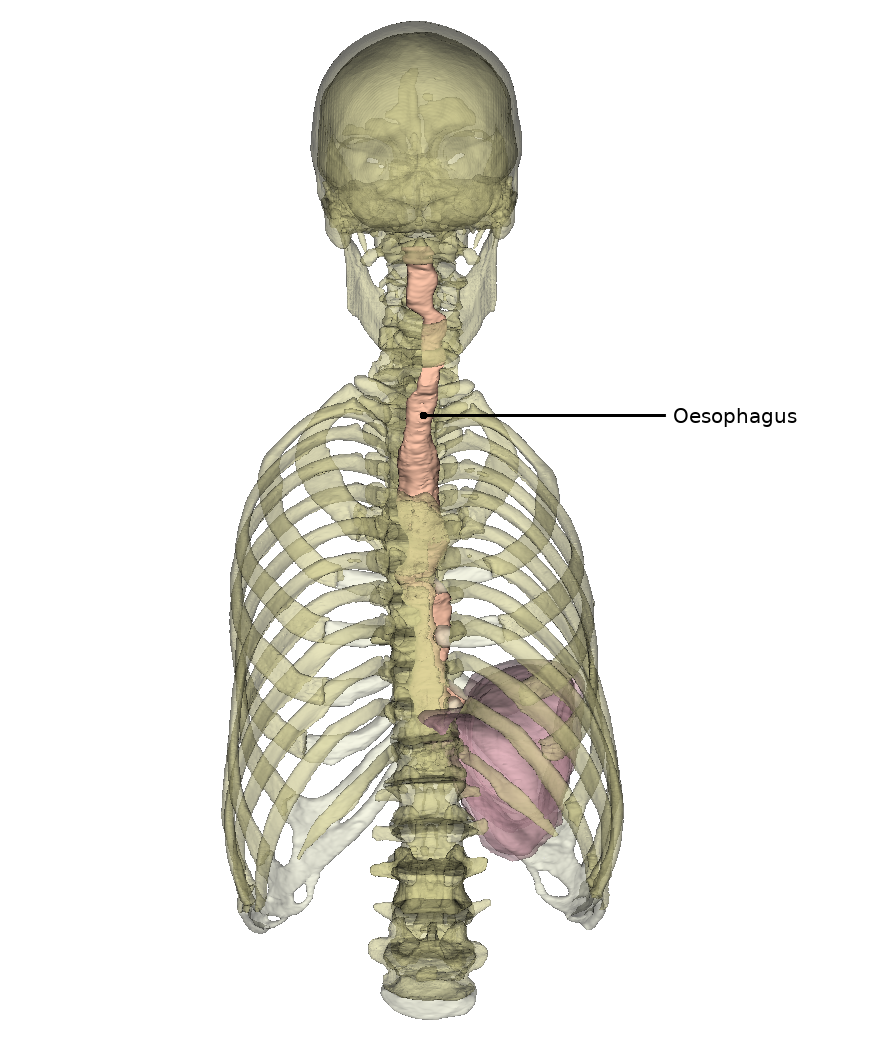
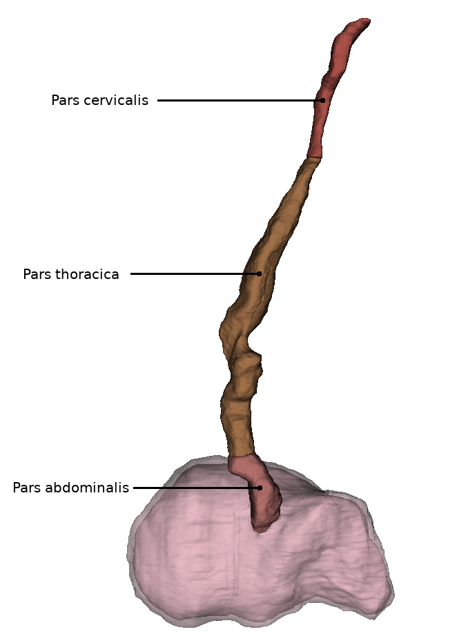
The human esophagus is approximately 23-27 centimeters long and has a diameter of 1-2 centimeters. It can be divided into three sections:
The pars cervicalis extends from HWK 6 to BWK 1 and lies anterior to the spine in the region of the neck.
The pars thoracica is the longest section and is located in the upper and posterior mediastinum (mediastinum superius and mediastinum posterius). It runs from BWK 1 to about BWK 11, where the passage through the diaphragm is located.
The shortest section is the pars abdominalis. This extends from the piercing of the diaphragm to the entrance of the stomach (lat. Cardia). In addition, it is already in the peritoneal cavity (lat. Cavitas peritonealis).
Projection onto the trunk
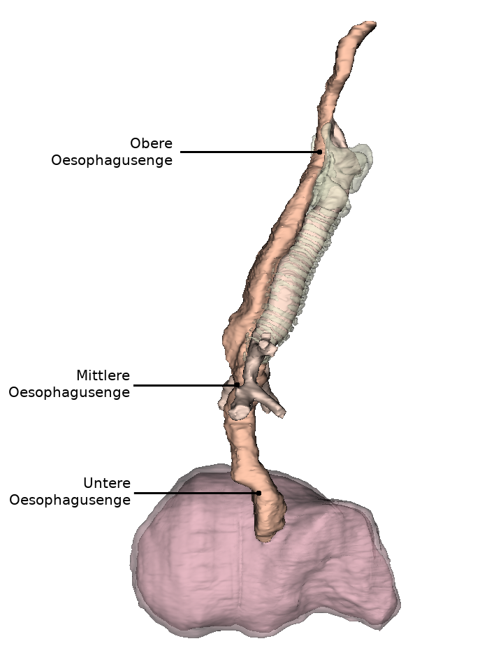
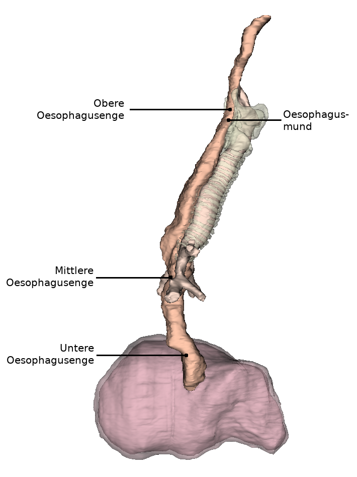
Due to its close relationship with neighboring organs, the esophagus has three narrowing points.
The esophageal orifice refers to the uppermost and narrowest part (constrictio pharyngooesophagealis). This is located in the pars cervicalis at the level of the cricoid cartilage of the larynx and measures approximately 14 millimeters in maximum diameter. Starting from the cricoid cartilage of the larynx, muscles surround the esophageal orifice and close it in the form of a sphincter, which is closed at rest.
The middle constriction (Constrictio partis thoracicae) in the pars thoracica is also called aortic stenosis. This is caused by the constriction of the esophagus by the left main bronchus and the aortic arch in the area of BWK 4/5. The maximum diameter here is 14 millimeters.
The inferior constriction (constrictio phrenica) results from the esophagus passing through the diaphragm at the beginning of the pars abdominalis and is called diaphragmatic constriction. Functional closure of the esophagus is ensured by the adjacent longitudinal tension of the musculature and by the veins of the esophageal wall. At rest (outside the act of swallowing), the lower part of the esophagus (pars abdominalis) is permanently closed.
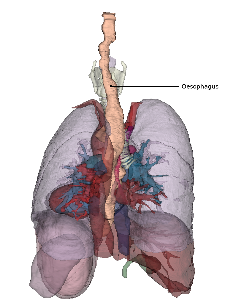
Position in horizontal section
In the ventral view of the thorax, the esophagus lies predominantly in the lower part and is slightly displaced to the right with respect to the median line by the aorta running to its left. A little below the Proc. xiphoideus sterni it enters the peritoneal cavity through the diaphragm.
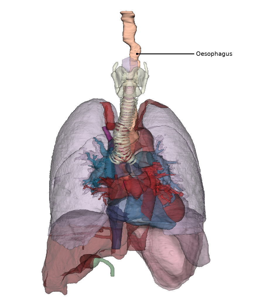
Relationship to neighbouring organs
Immediately on the anterior side of the esophagus runs the trachea until it divides into the two main bronchi (Latin bifurcatio tracheae). The anterior wall of the esophagus is connected to the posterior wall of the trachea by a multitude of connective tissue reins.
Below the structure of the tracheal bifurcation, in the lower mediastinum (Latin mediastinum inferius), the esophagus lies close to the left atrium and the thoracic aorta. Furthermore, the aorta initially runs caudally to the left of the esophagus and comes to rest behind the esophagus shortly before it enters the diaphragm.
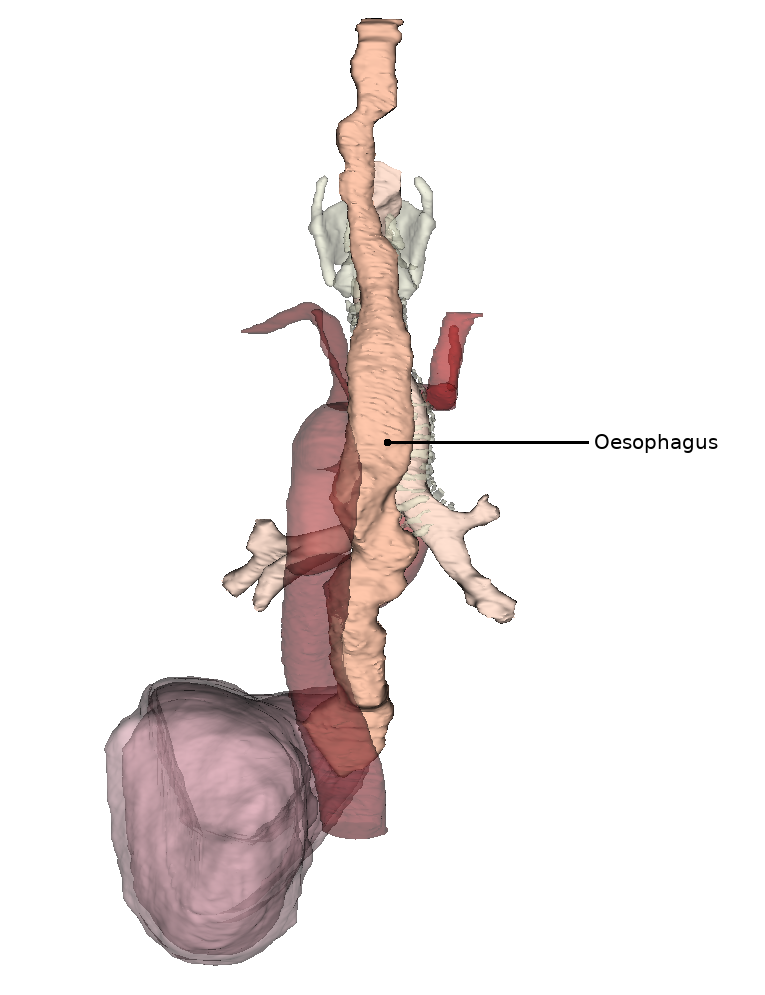
Position In situ
The esophagus has characteristic curvatures in addition to strictures.
The upper curvature runs in the view from ventral in the pars cervicalis to the left. The middle curvature is in the pars thoracica and runs to the right due to the nearby thoracic aorta. The inferior curvature in the pars abdominalis now turns back to the left.
If the esophagus is viewed in the sagittal plane, it can be seen that it follows the course of the spine and is curved forward in a slightly concave manner.
Diseases