Elle/en: Unterschied zwischen den Versionen
(Die Seite wurde neu angelegt: „<div class="clear"></div> ---- </div> <div class="button_style"> <ul>'''Subsequent structures'''<li class="mw-ui-button button_new">Special:MyLanguage/Oberar…“) |
Becher (Diskussion | Beiträge) |
||
| (57 dazwischenliegende Versionen von 4 Benutzern werden nicht angezeigt) | |||
| Zeile 1: | Zeile 1: | ||
| − | Together with the [[Special:MyLanguage/Speiche| | + | Together with the [[Special:MyLanguage/Speiche|radius]] the ulna forms the bones of the forearm. It is a typical long, straight tubular bone and lies on the side of the little finger in the arm. |
| − | < | + | {{ArticleMenu en|Links Übungsaufgaben=[[Special:MyLanguage/Übungsaufgaben: Obere Extremität|Upper extremity]][[Special:MyLanguage/Übungsaufgabe: Unterarmknochen|Forearm bone]][[Special:MyLanguage/Übungsaufgabe: Elle|Elle]]| |
| − | + | Segmentereinbettung=<segmenter-embedding public wsemb-id="UlnaFrau" file="UlnaFrau.seg" height="300" width="400"/>| | |
| − | + | Links Benachbarte Strukturen=[[Special:MyLanguage/Oberarmknochen|Humerus]][[Special:MyLanguage/Speiche|Spoke]][[Special:MyLanguage/Handknochen|Hand Bones]][[Special:MyLanguage/Handwurzelknochen|CARPAL BONE]]| | |
| + | Links Körperregionen=[[Special:MyLanguage/Unterarmknochen|Forearm bone]][[Special:MyLanguage/Knochen Obere Extremität|BONES UPPER EXTREMITY]][[Special:MyLanguage/Obere Extremität|UPPER EXTREMITY]]| | ||
| + | Links Organsystem=[[Special:MyLanguage/Röhrenknochen|Tubular bone]][[Special:MyLanguage/Knochen|Bones]][[Special:MyLanguage/Passiver Bewegungsapparat|Passive musculoskeletal system]][[Special:MyLanguage/Bewegungsapparat|Musculoskeletal system]]}} | ||
---- | ---- | ||
| − | + | ==Anatomy== | |
| − | |||
| − | |||
| − | < | + | {{ArticleGallery| |
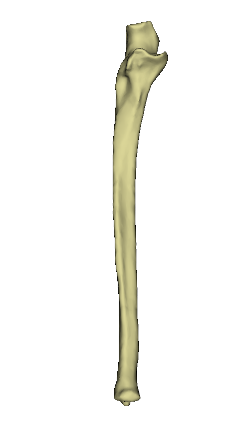
| − | -- | + | Bild 1=<lightbox-embedding src="file:Ulna.png" group="image-group-1" caption="View of the ulna (ventral)" width="400" height="300" style=""/>| |
| − | + | Weitere Bilder=<lightbox-embedding src="file:Ulna2.png" group="image-group-1" caption="View of the ulna (dorsal)"/> <lightbox-embedding src="file:Ulna4.png" group="image-group-1" caption="Ansicht der Ulna (distal)"/><lightbox-embedding src="file:Ulna3.png" group="image-group-1" caption="View of the ulna (proximal)"/> | |
| − | + | |title=Views of the ulna}} | |
| − | |||
| − | |||
| − | |||
| − | |||
| − | |||
| − | |||
| − | = | ||
| − | |||
| − | |||
| − | |||
| − | |||
| − | |||
| − | |||
| − | |||
| − | |||
| − | |||
| − | |||
| − | |||
| − | + | {{ArticleGallery| | |
| − | + | Bild 1=<lightbox-embedding src="file:UlnaVentral.png" group="image-group-1" caption="Anatomy ulna (ventral)" width="400" height="300" style=""/>| | |
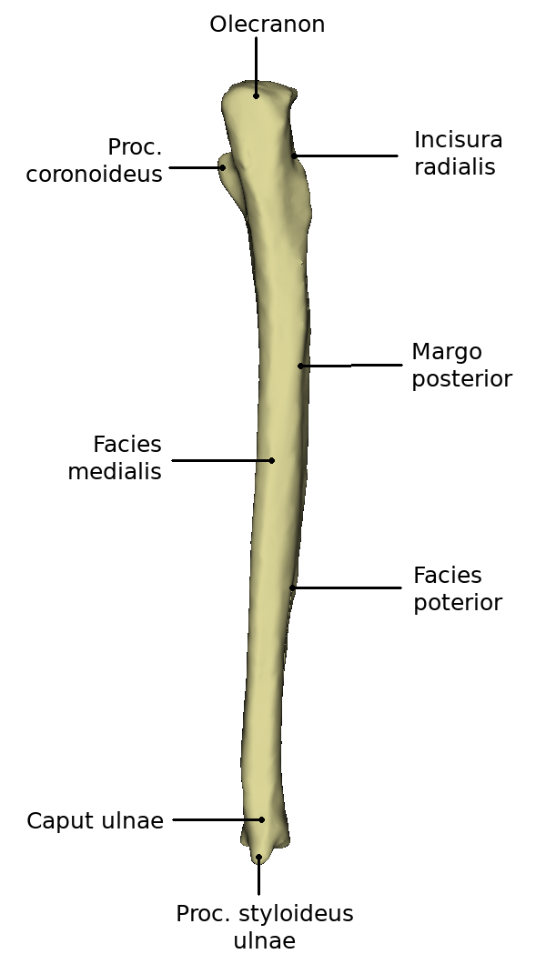
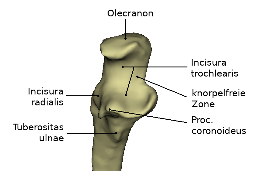
| − | <lightbox-embedding src="file:UlnaVentral.png" group="image-group-1" caption=" | + | Weitere Bilder=<lightbox-embedding src="file:UlnaDorsal.png" group="image-group-1" caption="Anatomy of the ulna (dorsal)"/><lightbox-embedding src="file:UlnaDistaleGelenkfläche.png" group="image-group-1" caption="Anatomy of the distal joint surface"/> <lightbox-embedding src="file:ProximaleGlenkflächenUlna.png" group="image-group-1" caption="Anatomy of the proximal joint surface"/> |
| − | + | |title=Anatomy of the ulna}} | |
| − | |||
| − | <lightbox-embedding src="file:UlnaDorsal.png" group="image-group-1" caption=" | ||
| − | |||
| − | |||
| − | |||
| − | |||
| − | + | The ulna has a corpus and one extremitas proximalis and distalis respectively. The Extremitas proximalis has a hook-shaped, roughened appendix, the Olecranon. In front is the Incisura trochlearis, which reaches up to the Processus coronoideus. Lateral is the Incisura radialis, into which the Circumferentia articularis (radii) is fitted. | |
| − | The ulna has a | ||
| − | |||
| − | === | + | === Proximal end=== |
| − | + | At the proximal extremity there is the hook-shaped bent extension, the olecranon. This has a certain roughness. In the front is the incisura trochlearis, which reaches up to the processus coronoideus. This depression is crescent-shaped and resembles a pair of forceps. Dorsally of these structures a roughened surface is found on the ulna. This is the attachment surface of the M. brachialis, the tuberosity ulnae. Lateral is the incisura radialis, into which the circumferentia articularis (radii) is fitted. | |
| − | ===Ulna- | + | ===Ulna-Shaft=== |
| − | + | The shaft of the ulna is also called the corpus ulnae. The cross-section of this structure has a triangular shape. Thus along the Corpus ulnae there are three sides and edges. The Margo interosseus represents the side facing the radius. This is where the Membrana interossea antebrachii, which connects ulna and radius, comes in. Furthermore, this edge serves as separation of the posterior and anterior facies. At the posterior side of the ulna the Margo posterior runs. This runs from the olecranon down to the proc. styloideus at the caput ulnae. Adjacent to this edge are the posterior and medial facies. | |
| − | + | The medial surface does not have a continuous uniform shape. At the proximal end, the surface is significantly wider than at the distal end. The facies posterior also changes its structure over time. It has a broad and concavely curved part at the end close to the body. This changes in the middle to a narrower, convex surface. The distal end of this surface is then rounded and flat. | |
| − | ===Ulna- | + | ===Ulna-head=== |
| − | + | The ulnar head or caput ulnae has the proc. styloideus ulnae at its distal end. This is also called the styloid process and is located on the small-fingered side of the distal joint surface. The distal end is generally widened in contrast to the shaft running in front of it. | |
| − | == | + | ==Function== |
| − | + | The ulna forms a functional unit with the radius. It creates the connection between elbow and wrist. The possibility of crossing the two bones allows the rotation of the forearm and the hand. | |
| − | == | + | ==Development== |
| − | + | The corpus ulnae begins to ossify already in the seventh week of embryonic development. Closure of the epiphyseal fugues begins between the fourth and seventh year of life. | |
| − | == | + | ==Diseases== |
| − | * [[Special:MyLanguage/Ulnafraktur| | + | * [[Special:MyLanguage/Ulnafraktur|Fracture of the ulna]] |
| − | == | + | ==Free exploration== |
| − | <div style="float:left;margin-right:1em;"><segmenter-embedding | + | <div style="float:left;margin-right:1em;"><segmenter-embedding public wsemb-id="UlnaFrau" file="UlnaFrau.seg" height="300" width="400"/></div> |
| − | <div style="float:left;width:50%"> | + | <div style="float:left;width:50%">Look at the structure of the ulna in 3D and explore it freely. Afterwards you can test your acquired knowledge by the exercises.</div> |
<div class="clear"></div> | <div class="clear"></div> | ||
---- | ---- | ||
| − | + | {{ArticleMenuEnd| | |
| − | + | Weiterer Artikel 1=[[Special:MyLanguage/Fibula|Fibula]]| | |
| − | + | Weiterer Artikel 2=[[Special:MyLanguage/Schlüsselbein|Collarbone]]}} | |
| − | + | ---- | |
| − | + | ||
| − | + | [[Category:Bones Upper Extremity]] | |
| − | + | [[Category:Upper Extremity]] | |
| − | + | [[Category:Body regions]] | |
| − | |||
| − | |||
<languages/> | <languages/> | ||
Aktuelle Version vom 10. Mai 2022, 12:23 Uhr
Together with the radius the ulna forms the bones of the forearm. It is a typical long, straight tubular bone and lies on the side of the little finger in the arm.
Inhaltsverzeichnis
Anatomy


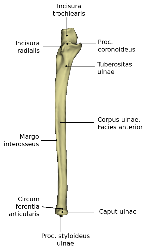

The ulna has a corpus and one extremitas proximalis and distalis respectively. The Extremitas proximalis has a hook-shaped, roughened appendix, the Olecranon. In front is the Incisura trochlearis, which reaches up to the Processus coronoideus. Lateral is the Incisura radialis, into which the Circumferentia articularis (radii) is fitted.
Proximal end
At the proximal extremity there is the hook-shaped bent extension, the olecranon. This has a certain roughness. In the front is the incisura trochlearis, which reaches up to the processus coronoideus. This depression is crescent-shaped and resembles a pair of forceps. Dorsally of these structures a roughened surface is found on the ulna. This is the attachment surface of the M. brachialis, the tuberosity ulnae. Lateral is the incisura radialis, into which the circumferentia articularis (radii) is fitted.
Ulna-Shaft
The shaft of the ulna is also called the corpus ulnae. The cross-section of this structure has a triangular shape. Thus along the Corpus ulnae there are three sides and edges. The Margo interosseus represents the side facing the radius. This is where the Membrana interossea antebrachii, which connects ulna and radius, comes in. Furthermore, this edge serves as separation of the posterior and anterior facies. At the posterior side of the ulna the Margo posterior runs. This runs from the olecranon down to the proc. styloideus at the caput ulnae. Adjacent to this edge are the posterior and medial facies. The medial surface does not have a continuous uniform shape. At the proximal end, the surface is significantly wider than at the distal end. The facies posterior also changes its structure over time. It has a broad and concavely curved part at the end close to the body. This changes in the middle to a narrower, convex surface. The distal end of this surface is then rounded and flat.
Ulna-head
The ulnar head or caput ulnae has the proc. styloideus ulnae at its distal end. This is also called the styloid process and is located on the small-fingered side of the distal joint surface. The distal end is generally widened in contrast to the shaft running in front of it.
Function
The ulna forms a functional unit with the radius. It creates the connection between elbow and wrist. The possibility of crossing the two bones allows the rotation of the forearm and the hand.
Development
The corpus ulnae begins to ossify already in the seventh week of embryonic development. Closure of the epiphyseal fugues begins between the fourth and seventh year of life.