Oberarmknochen/en: Unterschied zwischen den Versionen
(Die Seite wurde neu angelegt: „The humerus is articulately connected to shoulder blade, ulna and Special:MyLanguage/Speiche…“) |
(Die Seite wurde neu angelegt: „It consists of the corpus and a proximal and distal extremity. The proximal end forms the caput humeri with the adjacent neck, collum humeri. It appears slight…“) |
||
| Zeile 46: | Zeile 46: | ||
The humerus is articulately connected to [[Special:MyLanguage/Schulterblatt|shoulder blade]], [[Special:MyLanguage/Elle|ulna]] and [[Special:MyLanguage/Speiche|radius]]. | The humerus is articulately connected to [[Special:MyLanguage/Schulterblatt|shoulder blade]], [[Special:MyLanguage/Elle|ulna]] and [[Special:MyLanguage/Speiche|radius]]. | ||
| − | + | It consists of the corpus and a proximal and distal extremity. The proximal end forms the caput humeri with the adjacent neck, collum humeri. It appears slightly retracted and is also called Collum anatomicum. The distal end is formed by the condyle humeri.<br> | |
| − | + | At the anterior surface of the proximal extremity, the tubercle majus is located laterally and the tubercle minus medially. Between the two, there is a strong depression, the sulcus intertubercularis, which serves as a sliding rail for the tendon of the long caput of the biceps brachii.<br> | |
| − | + | At the posterior surface of the corpus humeri lies the sulcus n. radialis. At the distal extremity there is the medially more powerful bony protrusion Epicondylus medialis, laterally the weaker Epicondylus lateralis.<br> | |
| − | Trochlea humeri | + | Trochlea humeri and Capitulum humeri form the Condylus humeri and serve as the joint connection with the forearm bones. Medially from the trochlea humeri runs a shallow channel, the sulcus n. ulnaris. On the posterior surface above the trochlea there is a deep pit, the olecranon fossa. |
| − | + | The humerus is torqued at its proximal end, i.e. the caput is twisted about 20° backwards in relation to the axis through the epicondyli humeri (torsion). Between the longitudinal axis of the humerus and the caput there is an angle of about 130° on average, while at the distal end, between the transverse joint axis and the longitudinal axis of the humeral shaft, there is an angle of 76 to 89°.<br>. | |
| − | + | The bony socket (Cavitas glenoidalis) of the Articulatio humeri, a ball-and-socket joint, is considerably smaller than the humeral head. The socket is enlarged by a fibrocartilaginous joint lip, labrum glenoidale. The surface area of the cavitas glenoidalis is approximately 6 cm². The weight of the upper extremity is approximately 4 kg. Since there are no stronger ligaments, the muscles that surround the joint must secure it. This is therefore called a muscle-secured joint. The so-called "rotator cuff" is part of this muscular support and strengthens the joint capsule in particular. | |
| − | + | The caput humeri is approximately spherical. The synovial joint capsule is attached to the labrum glenoidale of the scapula. The synovial joint capsule is attached to the labrum glenoidale of the scapula and extends along the intracapsular tendon of the long head of the biceps like a sack, enclosing it with a vagina synovialis intertubercularis, which is located in the sulcus intertubercularis. | |
==Funktion== | ==Funktion== | ||
Version vom 26. Mai 2020, 11:40 Uhr
The upper arm bone (lat. Humerus) is a strong, straight and long tubular bone. It is the largest bone in the human arm. It forms the connection between shoulder and elbow. In general, the humerus is divided into the proximal end piece, the humeral shaft and the distal end piece.
- Subsequent structures
- Superordinate structures
Anatomy


The humerus is articulately connected to shoulder blade, ulna and radius.
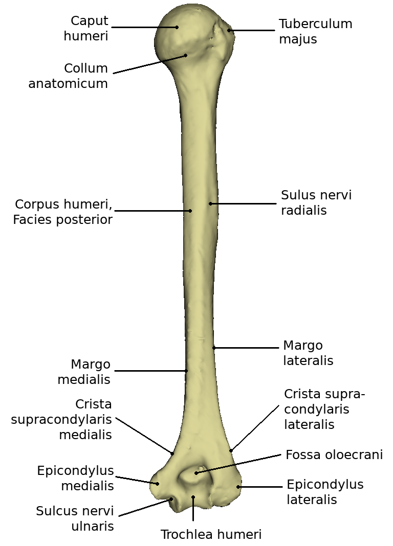
It consists of the corpus and a proximal and distal extremity. The proximal end forms the caput humeri with the adjacent neck, collum humeri. It appears slightly retracted and is also called Collum anatomicum. The distal end is formed by the condyle humeri.
At the anterior surface of the proximal extremity, the tubercle majus is located laterally and the tubercle minus medially. Between the two, there is a strong depression, the sulcus intertubercularis, which serves as a sliding rail for the tendon of the long caput of the biceps brachii.
At the posterior surface of the corpus humeri lies the sulcus n. radialis. At the distal extremity there is the medially more powerful bony protrusion Epicondylus medialis, laterally the weaker Epicondylus lateralis.
Trochlea humeri and Capitulum humeri form the Condylus humeri and serve as the joint connection with the forearm bones. Medially from the trochlea humeri runs a shallow channel, the sulcus n. ulnaris. On the posterior surface above the trochlea there is a deep pit, the olecranon fossa.
The humerus is torqued at its proximal end, i.e. the caput is twisted about 20° backwards in relation to the axis through the epicondyli humeri (torsion). Between the longitudinal axis of the humerus and the caput there is an angle of about 130° on average, while at the distal end, between the transverse joint axis and the longitudinal axis of the humeral shaft, there is an angle of 76 to 89°.
.
The bony socket (Cavitas glenoidalis) of the Articulatio humeri, a ball-and-socket joint, is considerably smaller than the humeral head. The socket is enlarged by a fibrocartilaginous joint lip, labrum glenoidale. The surface area of the cavitas glenoidalis is approximately 6 cm². The weight of the upper extremity is approximately 4 kg. Since there are no stronger ligaments, the muscles that surround the joint must secure it. This is therefore called a muscle-secured joint. The so-called "rotator cuff" is part of this muscular support and strengthens the joint capsule in particular.
The caput humeri is approximately spherical. The synovial joint capsule is attached to the labrum glenoidale of the scapula. The synovial joint capsule is attached to the labrum glenoidale of the scapula and extends along the intracapsular tendon of the long head of the biceps like a sack, enclosing it with a vagina synovialis intertubercularis, which is located in the sulcus intertubercularis.
Funktion
Der Humerus verbindet Schulter und Unterarm miteinander. Er ist an der Bildung der Schultergelenkes und des Elenbogengelenkes beteiligt. Der Oberarmknochen dient als Ansatz und Ursprung vieler Muskeln, die zur Bewegung des Armes und der Schulter benötigt werden.
Bewegung
Bewegungen in drei Freiheitsgraden sind möglich. Man spricht von Abduktion und Adduktion, wobei man von der Ruhestellung des Caput humeri in der Scapula-Ebene ausgeht. Man kennt die Anteversion, das nach vorne Heben des Armes und ihre Gegenbewegung, die Retroversion. Durch eine rotatorische Komponente ergibt sich unter Mitwirkung der vorher genannten Bewegungen eine zusammengesetzte Bewegung, die Zirkumduktion oder das Kreisen, wobei der Arm praktisch einen Kegelmantel beschreibt.
Bei den Abduktionsbewegungen kommt es immer zu einer Mitbewegung der Scapula; eine exzessive Mitbewegung der Scapula tritt bei einer Abduktion über 90 Grad ein (Elevation).
Erkrankungen
Freies Explorieren
- Weitere Artikel:
- Weiterführende Links: