Spina bifida/en: Unterschied zwischen den Versionen
Becher (Diskussion | Beiträge) |
Becher (Diskussion | Beiträge) (Die Seite wurde neu angelegt: „Category:Wirbelsäule/en Category:Knochen Rumpf/en Category:Rumpf/en Category:Körperregionen/en“) |
||
| Zeile 71: | Zeile 71: | ||
---- | ---- | ||
| − | [[Category:Wirbelsäule]] | + | [[Category:Wirbelsäule/en]] |
| − | [[Category:Knochen Rumpf]] | + | [[Category:Knochen Rumpf/en]] |
| − | [[Category:Rumpf]] | + | [[Category:Rumpf/en]] |
| − | [[Category:Körperregionen]] | + | [[Category:Körperregionen/en]] |
</div> | </div> | ||
<languages/> | <languages/> | ||
Version vom 31. Mai 2020, 09:10 Uhr
Spina bifida, colloquially "open back", is a congenital malformation of the spine. The spinal cord is not completely enclosed by the vertebral arch and the spinal membranes can emerge from the spinal canal.
Inhaltsverzeichnis
Reason
Spina bifida is a neural tube defect. It develops approximately in the third to fourth week of pregnancy. A vertebral gap occurs when there is an occlusion disorder in the lower section of the neural tube. This means that the two curved parts of the vertebrae do not fuse. Thus, the bony ring of vertebral body and vertebral arch around the vertebral hole is not closed. This results in a gap in the vertebra. This can be so pronounced that the spinous processes are missing in one or more vertebrae. The vertebrae of the lumbar spine and sacrum are affected significantly more often than the thoracic and cervical spine.
Shapes
The open back can occur in two forms. In spina bifida occulta, the vertebral arches are split, but the spinal cord does not emerge from the opening and is therefore unsplit. In this form there are often no symptoms and it is usually discovered by chance through X-rays.
Spina bifida aperta, on the other hand, is more problematic. In spina bifida aperta, the spinal meninges and/or the spinal cord emerge from the spinal gap. This is usually manifested by an outwardly formed sac. This is usually filled with spinal fluid. This form of spina bifida can cause severe symptoms such as paralysis of the stomach and intestines, bladder emptying disorders or even loss of pain or sensitivity disorders. Spina bifida aperta is surgically corrected immediately after birth. In some clinics, this correction can already be performed prenatally.
Comparison split vertebra with healthy vertebra
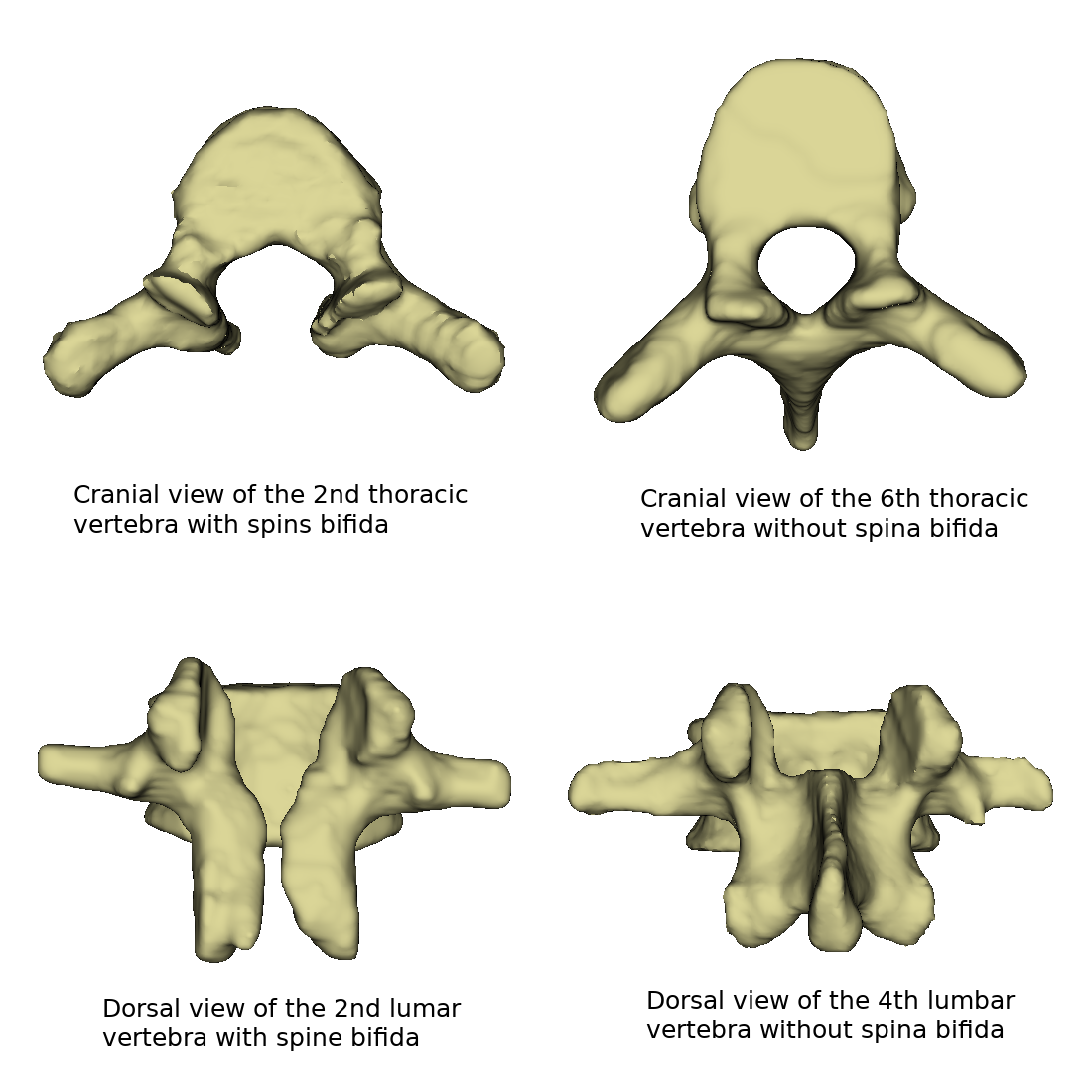
The upper pictures show one thoracic vertebra each. The left, second thoracic vertebra has spina bifida. This is so pronounced that there is no spinous process and the vertebral hole is wide open. For comparison, the sixth thoracic vertebra of the same spine is next to it. This one has a pronounced spinous process.
The second row shows one lumbar vertebra each. On the left, the second lumbar vertebra is shown with a vertebral gap. Next to it the fourth lumbar vertebra is shown. This is fully developed.
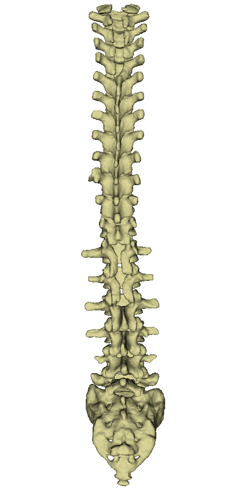
All these vertebrae originate from one spinal column (see upper figure). In this spine, there were split vertebrae in both the thoracic and lumbar regions. Furthermore, when imaging this spinal column, it should be noted that the cervical spine area is not completely shown. In the area between the lumbar and sacral vertebrae, there is also lumbalization. Thus, a sixth lumbar vertebra is visible in the spinal column.