Dataset:Patella Case 1/en: Unterschied zwischen den Versionen
Aus Dornheim Anatomy
Becher (Diskussion | Beiträge) (Die Seite wurde neu angelegt: „The dataset contains the processed 3D models of the patella of a female patient.“) |
Becher (Diskussion | Beiträge) (Die Seite wurde neu angelegt: „<h2>Bilder</h2> {{DatasetGallery| Bild 1=<lightbox-embedding src="file:Kniescheibe1.png" group="image-group-1" caption="View of the patella with the leg bones"…“) |
||
| Zeile 5: | Zeile 5: | ||
<h2>Bilder</h2> | <h2>Bilder</h2> | ||
{{DatasetGallery| | {{DatasetGallery| | ||
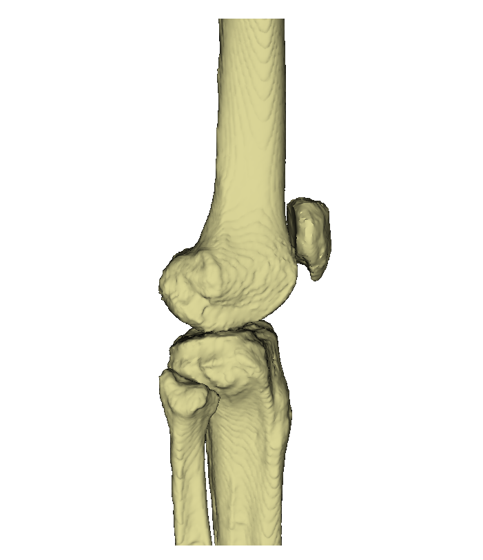
| − | Bild 1=<lightbox-embedding src="file:Kniescheibe1.png" group="image-group-1" caption=" | + | Bild 1=<lightbox-embedding src="file:Kniescheibe1.png" group="image-group-1" caption="View of the patella with the leg bones" width="300" height="300" style="width:300px;" />| |
| − | Bild 2=<lightbox-embedding src="file:Kniescheibe2.png" group="image-group-1" caption=" | + | Bild 2=<lightbox-embedding src="file:Kniescheibe2.png" group="image-group-1" caption="Patella (dorsal)" width="300" height="300" style="" />| |
| − | Bild 3=<lightbox-embedding src="file:Kniescheibe3.png" group="image-group-1" caption=" | + | Bild 3=<lightbox-embedding src="file:Kniescheibe3.png" group="image-group-1" caption="Patella (caudal)" width="300" height="300" />| |
| − | erneut Bild 2=<lightbox-embedding src="file:Kniescheibe2.png" group="image-group-1" caption=" | + | erneut Bild 2=<lightbox-embedding src="file:Kniescheibe2.png" group="image-group-1" caption="Patella (dorsal)" width="300" height="300" style="" />| |
| − | erneut Bild 3=<lightbox-embedding src="file:Kniescheibe3.png" group="image-group-1" caption=" | + | erneut Bild 3=<lightbox-embedding src="file:Kniescheibe3.png" group="image-group-1" caption="Patella (caudal)" width="300" height="300" />| |
| − | Weitere Bilder=<lightbox-embedding src="file:KniescheibeBein.png" group="image-group-1" caption=" | + | Weitere Bilder=<lightbox-embedding src="file:KniescheibeBein.png" group="image-group-1" caption="Anatomy of the patella with the leg bones |
| − | <lightbox-embedding src="file:KniescheibeBeschriftet1.png" group="image-group-1" caption=" | + | " width="150" height="150" /> |
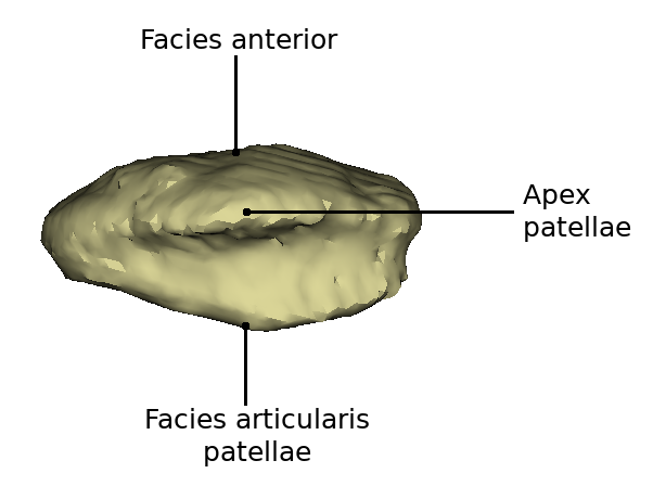
| − | <lightbox-embedding src="file:KniescheibeBeschriftet2.png" group="image-group-1" caption=" | + | <lightbox-embedding src="file:KniescheibeBeschriftet1.png" group="image-group-1" caption="Patella (ventral)" width="150" height="150" /> |
| − | <lightbox-embedding src="file:KniescheibeBeschriftet3.png" group="image-group-1" caption=" | + | <lightbox-embedding src="file:KniescheibeBeschriftet2.png" group="image-group-1" caption="Patella (dorsal)" width="150" height="150" /> |
| + | <lightbox-embedding src="file:KniescheibeBeschriftet3.png" group="image-group-1" caption="Patella (caudal)" width="150" height="150" /> | ||
|title=Datensatz Kniescheibe Fall 1}} | |title=Datensatz Kniescheibe Fall 1}} | ||
Version vom 6. Dezember 2021, 14:15 Uhr
Inhaltsverzeichnis
Dataset Patella Case 1
The dataset contains the processed 3D models of the patella of a female patient.
Bilder


Datensatz Kniescheibe Fall 1
Videos
Kniescheibe im CT-Datensatz
Die Kniescheibe im 3D-Modell
Wichtige anatomische Merkmale der Kniescheibe