Bauchspeicheldrüse/en: Unterschied zwischen den Versionen
Becher (Diskussion | Beiträge) |
(Übernehme Bearbeitung einer neuen Version der Quellseite) |
||
| Zeile 1: | Zeile 1: | ||
| − | <div class=" | + | <div class="mw-translate-fuzzy"> |
| − | |||
| − | |||
| − | |||
| − | |||
| − | |||
| − | |||
| − | |||
| − | |||
| − | |||
| − | |||
| − | |||
| − | |||
| − | |||
| − | |||
| − | |||
| − | |||
| − | |||
| − | |||
| − | |||
| − | |||
| − | |||
| − | |||
| − | |||
| − | |||
| − | |||
| − | |||
| − | |||
| − | |||
| − | |||
| − | |||
| − | |||
| − | |||
| − | |||
| − | |||
| − | |||
| − | |||
| − | |||
| − | |||
<div class="dropdown"> | <div class="dropdown"> | ||
<div class="floatright" style="margin:0.4em;">[[File:PfeilDropdown.png|20px|link=|Organ system]]</div> | <div class="floatright" style="margin:0.4em;">[[File:PfeilDropdown.png|20px|link=|Organ system]]</div> | ||
| Zeile 46: | Zeile 8: | ||
</div> | </div> | ||
---- | ---- | ||
| − | ==Structure of the pancreas== | + | ==Structure of the pancreas== |
| + | </div> | ||
The pancreas is a 14-18 cm long organ and has a weight of 70-80 g. It lies secondary retroperitoneally between stomach and abdominal aorta. The pancreas is surrounded by a thin connective tissue capsule (Capsula fibrosa), from which thin septa draw into the inner part of the gland. | The pancreas is a 14-18 cm long organ and has a weight of 70-80 g. It lies secondary retroperitoneally between stomach and abdominal aorta. The pancreas is surrounded by a thin connective tissue capsule (Capsula fibrosa), from which thin septa draw into the inner part of the gland. | ||
| Zeile 61: | Zeile 24: | ||
<!--{{MediaWiki-Button |Typ=progressive-normal |Link=Artikel |Text=Zum Artikel}}--> | <!--{{MediaWiki-Button |Typ=progressive-normal |Link=Artikel |Text=Zum Artikel}}--> | ||
| + | <div class="mw-translate-fuzzy"> | ||
==Projection of the pancreas onto the trunk== | ==Projection of the pancreas onto the trunk== | ||
<div class="thumb tright thumbinner"> | <div class="thumb tright thumbinner"> | ||
| Zeile 68: | Zeile 32: | ||
<div class="thumbcaption"> | <div class="thumbcaption"> | ||
Pancreas: Location of the pancreas</div> | Pancreas: Location of the pancreas</div> | ||
| − | </div> | + | </div> |
| + | </div> | ||
The pancreas lies transversely in the upper abdomen and mostly in the regio epigastrica. The location is called secondary retroperitoneal due to the change of position during embryonic time.<br> | The pancreas lies transversely in the upper abdomen and mostly in the regio epigastrica. The location is called secondary retroperitoneal due to the change of position during embryonic time.<br> | ||
| Zeile 78: | Zeile 43: | ||
==Pancreas in situ== | ==Pancreas in situ== | ||
| + | <div class="mw-translate-fuzzy"> | ||
<div class="thumb tright thumbinner"> | <div class="thumb tright thumbinner"> | ||
<div class="picture"> | <div class="picture"> | ||
| Zeile 84: | Zeile 50: | ||
<div class="thumbcaption" style="width:300px"> | <div class="thumbcaption" style="width:300px"> | ||
Pancreas: Horizontal section approximately at the height of 12th thoracic or 1st lumbar vertebral body.</div> | Pancreas: Horizontal section approximately at the height of 12th thoracic or 1st lumbar vertebral body.</div> | ||
| − | </div> | + | </div> |
| + | </div> | ||
| + | |||
Removed from the <!--[segmenter_snapshot pancreassitu 0]-->"WebViewer"<!--[/]-->: liver, stomach, parts of the large and small intestine. For a better overview of the structures in the vicinity of the pancreas, the retroperitoneal connective and adipose tissue as well as the capsula adiposa renis are not shown.<br> | Removed from the <!--[segmenter_snapshot pancreassitu 0]-->"WebViewer"<!--[/]-->: liver, stomach, parts of the large and small intestine. For a better overview of the structures in the vicinity of the pancreas, the retroperitoneal connective and adipose tissue as well as the capsula adiposa renis are not shown.<br> | ||
| Zeile 92: | Zeile 60: | ||
==Diseases== | ==Diseases== | ||
| + | <div class="mw-translate-fuzzy"> | ||
*[[Special:MyLanguage/Bauchspeicheldrüsenentzündung|Pancreatitis]] | *[[Special:MyLanguage/Bauchspeicheldrüsenentzündung|Pancreatitis]] | ||
*[[Special:MyLanguage/Pankreasinsuffizienz|Pancreatic insufficiency]] | *[[Special:MyLanguage/Pankreasinsuffizienz|Pancreatic insufficiency]] | ||
*[[Special:MyLanguage/Pankreaszysten|Pancreatic zyste]] | *[[Special:MyLanguage/Pankreaszysten|Pancreatic zyste]] | ||
| + | </div> | ||
==Free exploration== | ==Free exploration== | ||
| + | <div class="mw-translate-fuzzy"> | ||
{{Tab2|Button1=Pancreas|Button2=Pancreas in situ|Segmenter1=<segmenter-embedding public wsemb-id="Bauchspeicheldruese" file="Bauchspeicheldruese.seg" height="300" width="400"/>|Inhalt1=<b>Pancreas</b><br>View the pancreas in 3D and explore it freely. Afterwards, you can look at other cases or check your learned knowledge through the practice exercises.|Segmenter2=<segmenter-embedding public wsemb-id="Bauchspeicheldrueseinsitu" file="Bauchspeicheldrueseinsitu.seg" height="300" width="400"/>|Inhalt2=<b>Case 1: Pancreas in situ </b><br>View the pancreas in situ.}} | {{Tab2|Button1=Pancreas|Button2=Pancreas in situ|Segmenter1=<segmenter-embedding public wsemb-id="Bauchspeicheldruese" file="Bauchspeicheldruese.seg" height="300" width="400"/>|Inhalt1=<b>Pancreas</b><br>View the pancreas in 3D and explore it freely. Afterwards, you can look at other cases or check your learned knowledge through the practice exercises.|Segmenter2=<segmenter-embedding public wsemb-id="Bauchspeicheldrueseinsitu" file="Bauchspeicheldrueseinsitu.seg" height="300" width="400"/>|Inhalt2=<b>Case 1: Pancreas in situ </b><br>View the pancreas in situ.}} | ||
| + | </div> | ||
| + | <div class="mw-translate-fuzzy"> | ||
---- | ---- | ||
<div class="clear aufgaben" style="margin-bottom:1em;"> | <div class="clear aufgaben" style="margin-bottom:1em;"> | ||
| Zeile 106: | Zeile 79: | ||
<li class="mw-ui-button button_new" >[[Special:MyLanguage/Übungsaufgaben|Exercises]]</li> | <li class="mw-ui-button button_new" >[[Special:MyLanguage/Übungsaufgaben|Exercises]]</li> | ||
</div> | </div> | ||
| − | |||
| − | |||
| − | |||
| − | |||
| − | |||
| − | |||
| − | |||
| − | |||
| − | |||
| − | |||
| − | |||
| − | |||
| − | |||
</div> | </div> | ||
</div> | </div> | ||
| − | |||
| − | |||
| − | |||
| − | |||
| − | |||
| − | |||
</div> | </div> | ||
<languages/> | <languages/> | ||
| + | [[Category:Organe Abdomen]] | ||
| + | [[Category:Innere Organe]] | ||
| + | [[Category:Verdauungsapparat]] | ||
| + | [[Category:Organsystem]] | ||
| + | [[Category:Körperregionen]] | ||
Version vom 16. März 2022, 10:41 Uhr
Inhaltsverzeichnis
Structure of the pancreas
The pancreas is a 14-18 cm long organ and has a weight of 70-80 g. It lies secondary retroperitoneally between stomach and abdominal aorta. The pancreas is surrounded by a thin connective tissue capsule (Capsula fibrosa), from which thin septa draw into the inner part of the gland.
The pancreas is divided into three sections:
Pancreas head, body, and tail.
In addition, the pancreas has an excretory duct, the ductus panvreaticus and a common bile duct (ductus chloledochus). These two usually flow together on the papilla duodeni major into the pars descendens duodeni. The residue of the formerly dorsal pancreatic duct in the caput pancreatis, the ductus pancreaticus accessrius, flows into the papilla duodeni minor.
There are different variations of the duct:
- both ducts merge into one, which ends on a papilla (see "WebViewer")
- ducts remain separated from each other and end in two papillae
- In rare cases, in addition to the two points mentioned above, the ductus coledochus may lead seperately into the duodenum.
Projection of the pancreas onto the trunk

The pancreas lies transversely in the upper abdomen and mostly in the regio epigastrica. The location is called secondary retroperitoneal due to the change of position during embryonic time.
The caput pancreatis runs up to the 2/3 lumbar vertebra and the corpus pancreatis up to the 1/2 lumbar vertebra. The caput pancreatis can extend to the spleen and is located in the left upper abdomen.
Pancreas in situ
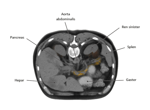
Removed from the "WebViewer": liver, stomach, parts of the large and small intestine. For a better overview of the structures in the vicinity of the pancreas, the retroperitoneal connective and adipose tissue as well as the capsula adiposa renis are not shown.
The pancreas is secondary retroperitoneally located at the posterior wall of the bursa omentalis.
Diseases
Free exploration
- Pancreas
- Pancreas in situ
View the pancreas in 3D and explore it freely. Afterwards, you can look at other cases or check your learned knowledge through the practice exercises.
View the pancreas in situ.