Schlüsselbein/en: Unterschied zwischen den Versionen
Becher (Diskussion | Beiträge) (Die Seite wurde neu angelegt: „==Evolution==“) |
Becher (Diskussion | Beiträge) (Die Seite wurde neu angelegt: „During embryonic development, the clavicle is developed from a connective tissue base. It is therefore the only bone in an extremity that is not preformed by c…“) |
||
| Zeile 49: | Zeile 49: | ||
==Evolution== | ==Evolution== | ||
| − | + | During embryonic development, the clavicle is developed from a connective tissue base. It is therefore the only bone in an extremity that is not preformed by cartilage. However, the clavicle is one of the bones that develops earliest. Its diaphysis thus ossifies as early as embryonic week six to seven. An ossified nucleus can only be detected in the epyphyses at the age of 16 to 18 years. The epiphysis joints here do not close until the age of 20 to 25 years. They are therefore the last joints to close in the human body. | |
Version vom 1. April 2020, 09:53 Uhr
The clavicle (lat. Clavicula) is a pairwise tubular bone. It has an "s"-shaped bend and is located in the shoulder girdle. It is the only direct connection between the free upper extremity and the trunk.
Inhaltsverzeichnis
- Superordinate structures:
- Adjoining structures:
Anatomy


The clavicle is located in the shoulder girdle and thus at the front of the trunk. Medially it is limited by the Sternum and laterally by the arcomion of the shoulder blade. The connection to the sternum is the only direct link between the free upper extremity and the trunk.
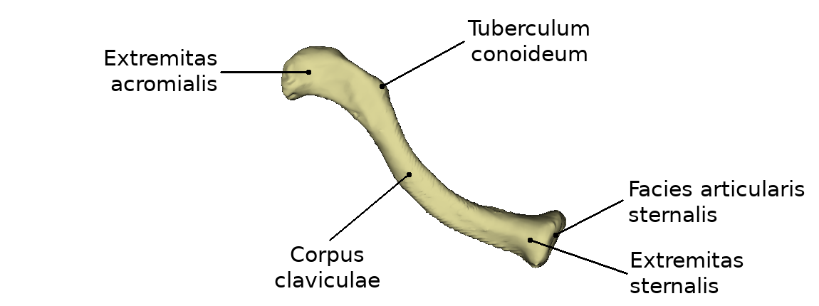
The clavicle is a tubular bone bent into an "S" shape. From cranial view the medial end shows a ventral convexity. This convexity takes up about 2/3 of the length of the entire bone. The lateral side, in contrast, shows a ventral concavity. In an adult, the clavicle is 12-15 cm long and can be palpated over its entire length.
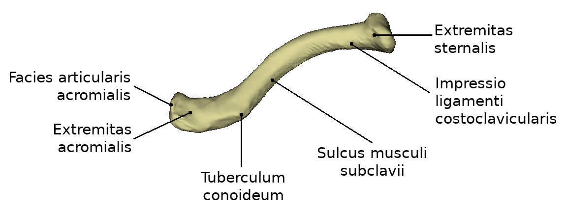
The thickened sternal end of the clavicle is called Extremitas sternalis. This name is due to its location adjacent to the sternum. On this side of the clavicle a saddle-shaped, three-sided articular surface is formed, which is covered with the Facies articularis sternalis. The lateral side of the clavicle is called Extremitas acromialis due to its connection to the acromion of the scapula. There is a rather flat, vertically oriented surface. This is almost oval and covered with the Facies articularis acrommialis. The area of the clavicle between the two ends is called Corpus claviculae.
If one looks at the clavicle from caudal, further structures become apparent. On the acromial side a bony elevation is pronounced, the tuberculum conoideum. This is also visible from the cranial view. Lateral to it is the linea trapezoides from the caudal side. From caudal view the corpus claviculae is characterized by an elongated depression called sulcus musculi subclavia. From the caudal side a depression appears at the sternal extremity, called impressio ligamenti costoclavicularis.
Joints
The collarbone is connected to the sternum via the sternoclavicular joint. This connects the following bony structures: Extremitas sternalis and Manubrium sterni.
The clavicle and the scapula articulate via the acromioclavicular joint. This connects the extremitas acromialis and the acromion of the scapula.
Evolution
During embryonic development, the clavicle is developed from a connective tissue base. It is therefore the only bone in an extremity that is not preformed by cartilage. However, the clavicle is one of the bones that develops earliest. Its diaphysis thus ossifies as early as embryonic week six to seven. An ossified nucleus can only be detected in the epyphyses at the age of 16 to 18 years. The epiphysis joints here do not close until the age of 20 to 25 years. They are therefore the last joints to close in the human body.
Erkrankungen
Freies Explorieren
further links