Schulterblatt/en: Unterschied zwischen den Versionen
(Die Seite wurde neu angelegt: „When viewed from ventral, the shoulder blade is characterized by a flat, slightly hollowed surface. This surface is the <i>Facies posterior</i>. The concave de…“) |
(Die Seite wurde neu angelegt: „From dorsal view the back of the scapula, the <i>Facies dorsalis</i> (also <i>Facies posterior</i>) is visible. It is divided into two parts by the spine of sc…“) |
||
| Zeile 84: | Zeile 84: | ||
When viewed from ventral, the shoulder blade is characterized by a flat, slightly hollowed surface. This surface is the <i>Facies posterior</i>. The concave depression is called <i>Fossa subscaolaris</i>. Under certain circumstances <i>Linea musculares</i> may be visible on this surface. | When viewed from ventral, the shoulder blade is characterized by a flat, slightly hollowed surface. This surface is the <i>Facies posterior</i>. The concave depression is called <i>Fossa subscaolaris</i>. Under certain circumstances <i>Linea musculares</i> may be visible on this surface. | ||
| − | + | From dorsal view the back of the scapula, the <i>Facies dorsalis</i> (also <i>Facies posterior</i>) is visible. It is divided into two parts by the spine of scapula, the <i>spina scapulae</i>. Thus the upper smaller part, the <i>Fossa supraspinata</i> and the lower larger part, the <i>Fossa infraspinata</i>, are formed. The <i>spina scapulae</i> begins at the inner edge and extends to the lateral shoulder angle. It increases in height during its course and ends in the acromion (shoulder height or acromion). This is a flattened bone process. At the acromion there is an oval joint surface, the <i>Facies articularis clavicularis</i>, which forms the connection to the clavicle through the acromioclavicular joint. The <i>angulus acromii</i> is formed at the transition of the lateral edge of the acromion to the scapular spine. This is an easily palpable bone point. | |
Die Gelenkpfanne (<i>Cavitas glenoidales</i>) befindet sich im <i>Angulus lateralis</i>. Dort artikulieren Scapula und Humerus miteinander. Am oberen Rand der Gelenkpfanne befindet sich ein knöchernes Höckerchen, dieser ist der sogenannte <i>Tuberculum supraglenoidale</i>. Unterhalb des <i>Cavitas glenoidales</i> sitzt das <i>Tuberculum infraglenoidale</i>. An die Gelenkpfanne schließt sich in Richtung der großen Knochenfläche der <i>Collum Scapulae</i>, der Hals der Scapula, an. Oberhalb der <i>Cavitas glenoidales</i> erstreckt sich der aus dem <i>Margo superior</i> entspringende Rabenschnabelfortsatz, der <i>Processus Coracoideus</i>. Er biegt sich rechtwinklig nach lateroventral um und endet abgeplattet. Das Acromion und der Rabenschnabelfortsatz dienen zusammen als Schutz des Gelenks. | Die Gelenkpfanne (<i>Cavitas glenoidales</i>) befindet sich im <i>Angulus lateralis</i>. Dort artikulieren Scapula und Humerus miteinander. Am oberen Rand der Gelenkpfanne befindet sich ein knöchernes Höckerchen, dieser ist der sogenannte <i>Tuberculum supraglenoidale</i>. Unterhalb des <i>Cavitas glenoidales</i> sitzt das <i>Tuberculum infraglenoidale</i>. An die Gelenkpfanne schließt sich in Richtung der großen Knochenfläche der <i>Collum Scapulae</i>, der Hals der Scapula, an. Oberhalb der <i>Cavitas glenoidales</i> erstreckt sich der aus dem <i>Margo superior</i> entspringende Rabenschnabelfortsatz, der <i>Processus Coracoideus</i>. Er biegt sich rechtwinklig nach lateroventral um und endet abgeplattet. Das Acromion und der Rabenschnabelfortsatz dienen zusammen als Schutz des Gelenks. | ||
Version vom 26. Mai 2020, 10:18 Uhr
The shoulder blade (lat. scapula) together with the Clavicula forms the bony Shoulder Belt. The shoulder blade represents a flat three-sided bone, which occurs in pairs, just like the clavicle. It is located on the dorsal thorax and is completely embedded in muscle loops. It has a connection to the Upper arm bone and to the clavicle.
Inhaltsverzeichnis
- Subsequent structures:
- Übergeordnete Strukturen:
Anatomy


The shoulder blade is a three-sided, flat bone. Together with the clavicle it forms the shoulder girdle. It has a variety of bony structures. The scapula is located dorsal to the thorax and is completely embedded in muscles. This musculature forms the connection to the Torso. On the shoulder blade, the shoulder joint is formed at the connection to the humerus. Via the Acromioclavicular joint the shoulder blade and clavicle articulate with each other.
In normal position, the scapula extends from the second to the seventh rib. The angulus inferior is located at the level of the spinous process of the seventh rib. The spina scapulae is located at the level of the spinous process of the third lumbar vertebra. In normal position, the scapula has a slight lateral rotation, so that the medial edge has an angle of about 3-5° to the spinous process.
Ränder

The flat bone is bordered by three edges. The upper edge is called margo superior. It represents the shortest edge of the bone and runs from the coracoid process (Proc. coracoideus) to the angulus superior. At this edge, medial to the base of the coracoid process, there is a distinct incision in the bony structure, the Incisura scapulae.
The Margo medialis is the edge directed medially, towards the middle of the back. This is the longest side of the shoulder blade. It is located between the Angulus superior and the Angulus inferior.
The outer margin ,Margo lateralis, points laterally. This margin is the most massive of the three. It begins at the lower edge of the glenoid cavity and extends to the lower shoulder blade angle (Angulus inferior).
Angle
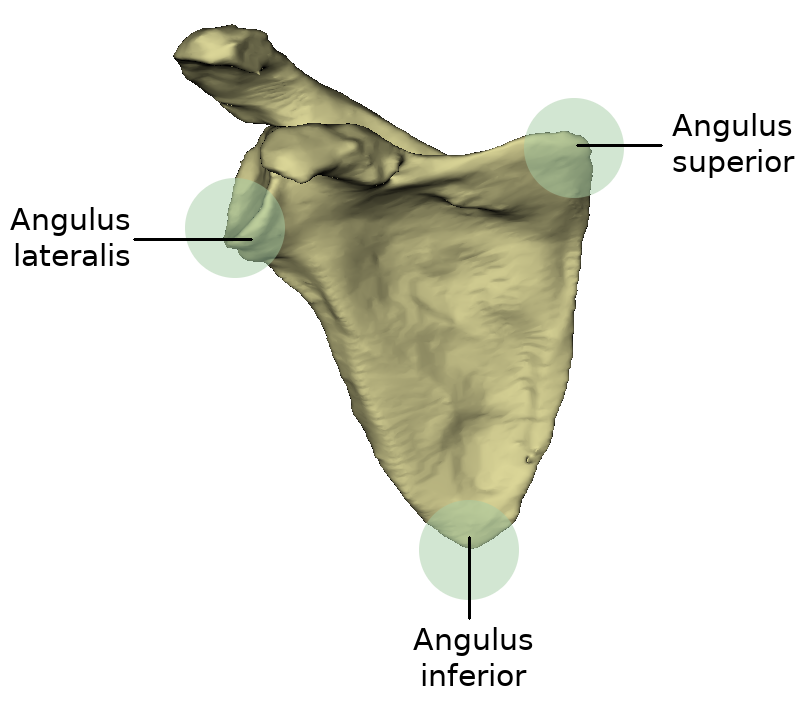
Because the shoulder blade resembles a triangle, three angles can be seen on it. Between the inner edge and the upper edge the angulus superior is enclosed. It represents the upper shoulder angle and is a rounded and flat bone area.
Between the Margo medialis and the outer edge (Margo lateralis) is the lower angle of the shoulder blade, the Angulus inferior. The origin of the Musculus teres major and some fibres of the Musculus latissimus dorsi is located at the rear surface of this angle.
The angulus lateralis is the lateral angle of the shoulder blade and is also called the "shoulder head" because it represents the most massive part of the scapula. It is located at the level of the cavitas glenoidalis.
Bony structure
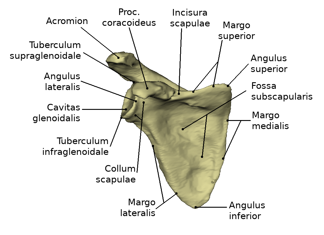

When viewed from ventral, the shoulder blade is characterized by a flat, slightly hollowed surface. This surface is the Facies posterior. The concave depression is called Fossa subscaolaris. Under certain circumstances Linea musculares may be visible on this surface.
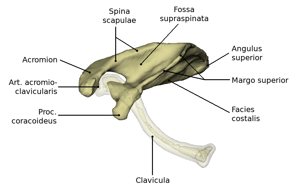
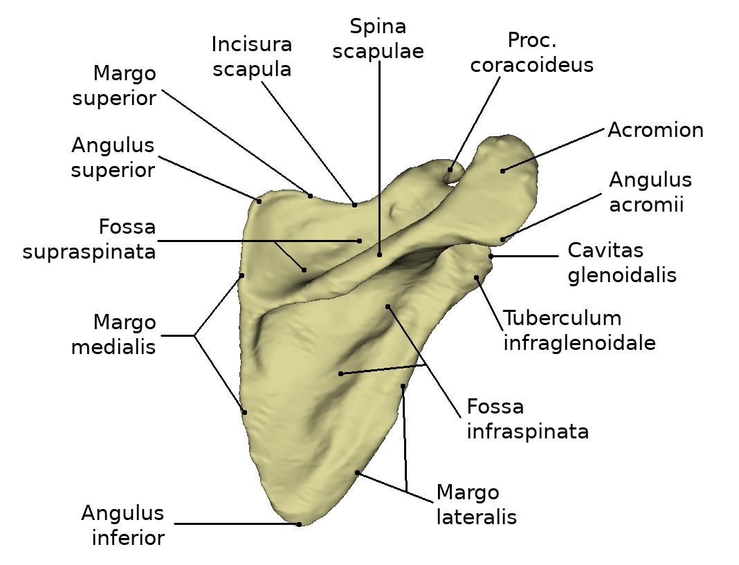
From dorsal view the back of the scapula, the Facies dorsalis (also Facies posterior) is visible. It is divided into two parts by the spine of scapula, the spina scapulae. Thus the upper smaller part, the Fossa supraspinata and the lower larger part, the Fossa infraspinata, are formed. The spina scapulae begins at the inner edge and extends to the lateral shoulder angle. It increases in height during its course and ends in the acromion (shoulder height or acromion). This is a flattened bone process. At the acromion there is an oval joint surface, the Facies articularis clavicularis, which forms the connection to the clavicle through the acromioclavicular joint. The angulus acromii is formed at the transition of the lateral edge of the acromion to the scapular spine. This is an easily palpable bone point.
Die Gelenkpfanne (Cavitas glenoidales) befindet sich im Angulus lateralis. Dort artikulieren Scapula und Humerus miteinander. Am oberen Rand der Gelenkpfanne befindet sich ein knöchernes Höckerchen, dieser ist der sogenannte Tuberculum supraglenoidale. Unterhalb des Cavitas glenoidales sitzt das Tuberculum infraglenoidale. An die Gelenkpfanne schließt sich in Richtung der großen Knochenfläche der Collum Scapulae, der Hals der Scapula, an. Oberhalb der Cavitas glenoidales erstreckt sich der aus dem Margo superior entspringende Rabenschnabelfortsatz, der Processus Coracoideus. Er biegt sich rechtwinklig nach lateroventral um und endet abgeplattet. Das Acromion und der Rabenschnabelfortsatz dienen zusammen als Schutz des Gelenks. Die knöcherne Gelenkpfanne Cavitas glenoidalis des Articulatio humeri, eines Kugelgelenkes, ist wesentlich kleiner als der Humeruskopf. Die Pfanne wird durch eine faserknorpelige Gelenklippe, Labrum glenoidale, vergrößert. Die Oberfläche der Cavitas glenoidalis beträgt ca. 6qcm. Das Gewicht der oberen Extremität beträgt etwa 4 kg. Da keine stärkeren Bänder vorhanden sind, müssen die Muskeln, die das Gelenk umhüllen, dieses sichern. Man spricht daher von einem muskelgesicherten Gelenk. Die sogenannte “Rotatorenmanschette” ist Teil dieser muskulären Sicherung und verstärkt im besonderen die Gelenkkapsel.
Bewegungen
Das Caput humeri ist annähernd kugelförmig. Die synoviale Gelenkkapsel ist am Labrum glenoidale der Scapula befestigt. Bewegungen sind in drei Freiheitsgraden möglich. Man spricht von Abduktion und Adduktion, wobei man von der Ruhestellung des Caput humeri in der Scapula-Ebene ausgeht. Man kennt die Anteversion, das nach vorne Heben des Armes, und ihre Gegenbewegung, die Retroversion. Durch eine rotatorische Komponente ergibt sich unter Mitwirkung der vorher genannten Bewegungen eine zusammengesetzte Bewegung, die Zirkumduktion oder das Kreisen, wobei der Arm praktisch einen Kegelmantel beschreibt. Bei den Abduktionsbewegungen kommt es immer zu einer Mitbewegung der Scapula; eine exzessive Mitbewegung der Scapula tritt bei einer Abduktion über 90 Grad ein (Elevation).
Erkrankungen
Freies Explorieren
- Weitere Artikel:
- Weiterführende Links: