Esophagus
The esophagus (Latin Oesophagus) is a muscular tube that, as part of the digestive system, carries swallowed food from the pharynx to the stomach by means of peristaltic movements.
Anatomy

Division
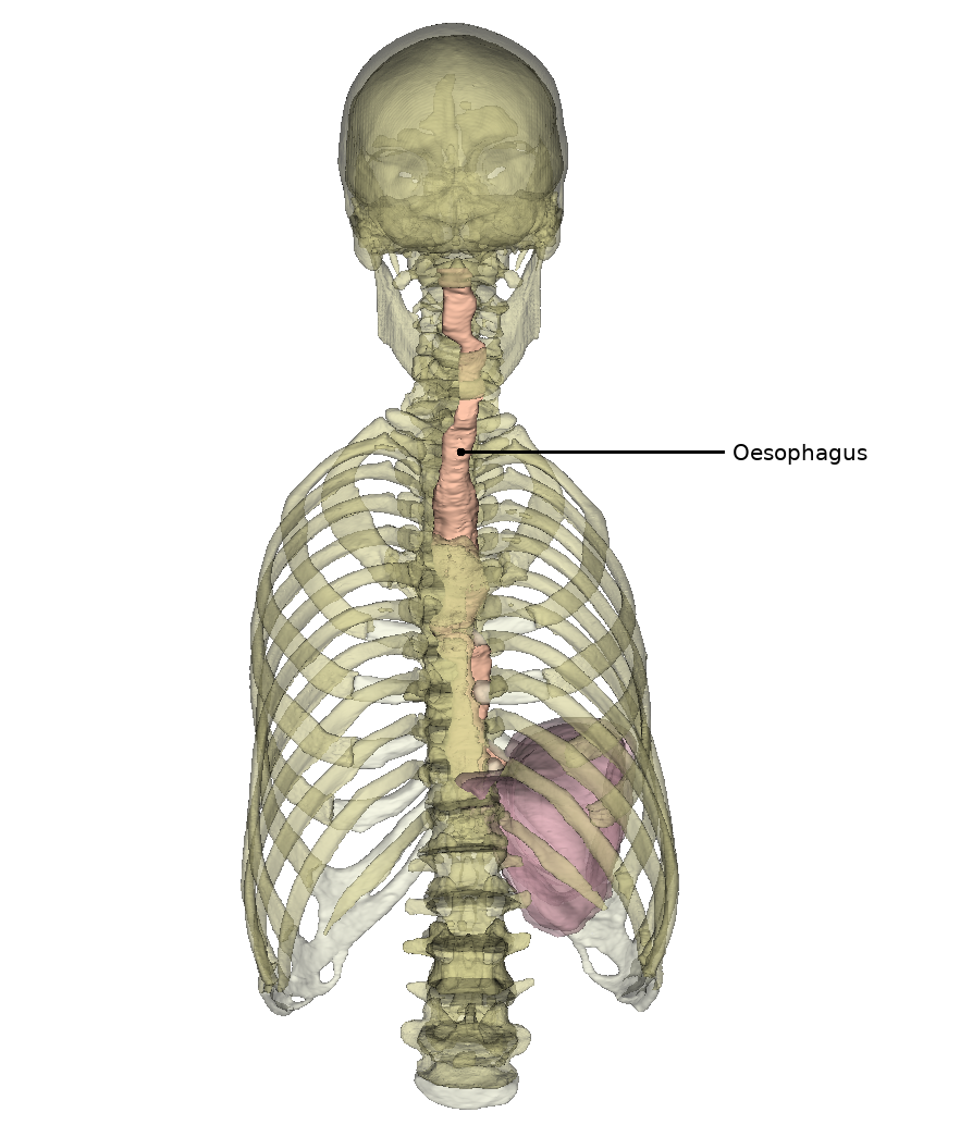
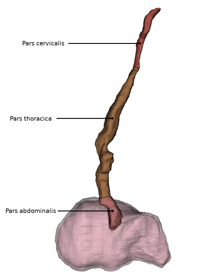
The human esophagus is approximately 23-27 centimeters long and has a diameter of 1-2 centimeters. It can be divided into three sections:
The pars cervicalis extends from HWK 6 to BWK 1 and lies anterior to the spine in the region of the neck.
The pars thoracica is the longest section and is located in the upper and posterior mediastinum (mediastinum superius and mediastinum posterius). It runs from BWK 1 to about BWK 11, where the passage through the diaphragm is located.
The shortest section is the pars abdominalis. This extends from the piercing of the diaphragm to the entrance of the stomach (lat. Cardia). In addition, it is already in the peritoneal cavity (lat. Cavitas peritonealis).
Projection onto the trunk
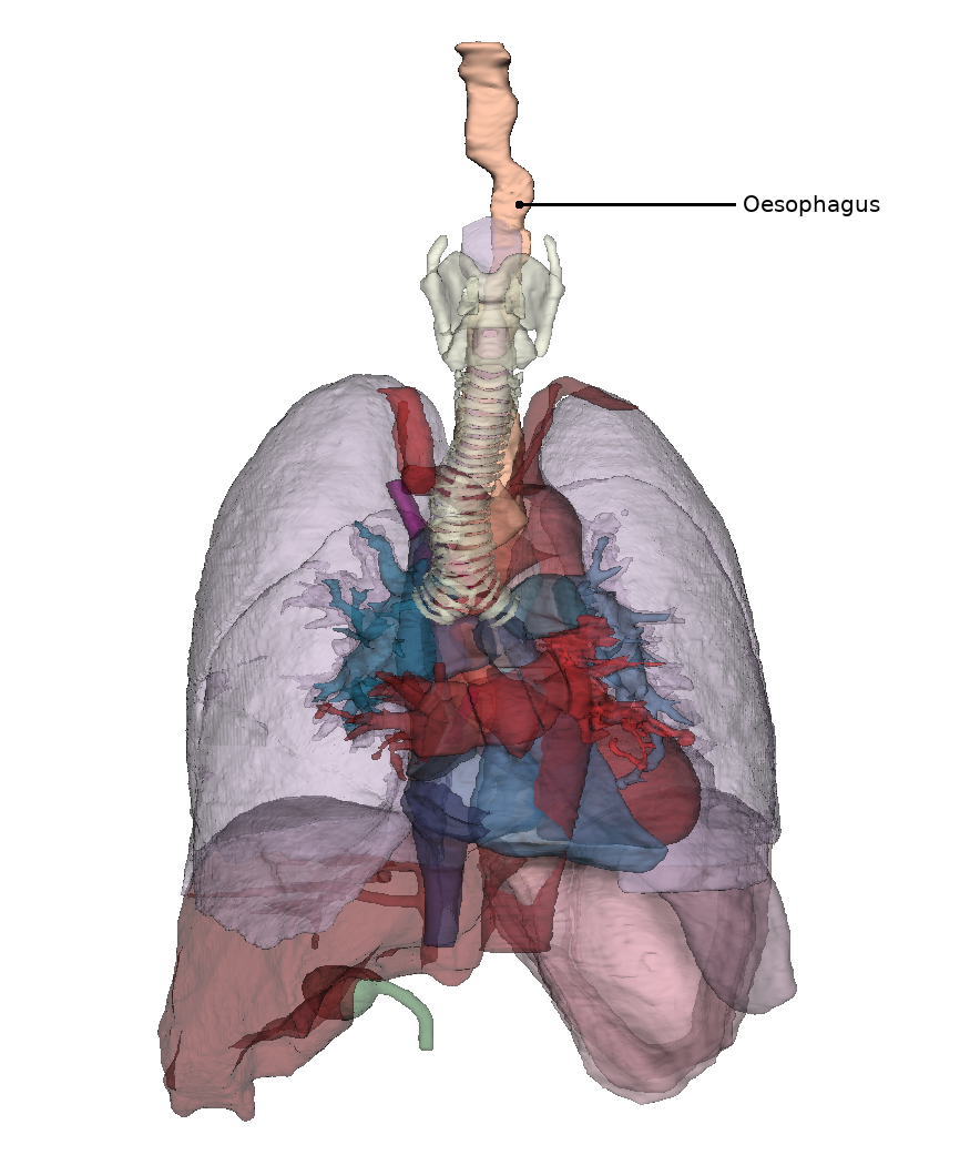
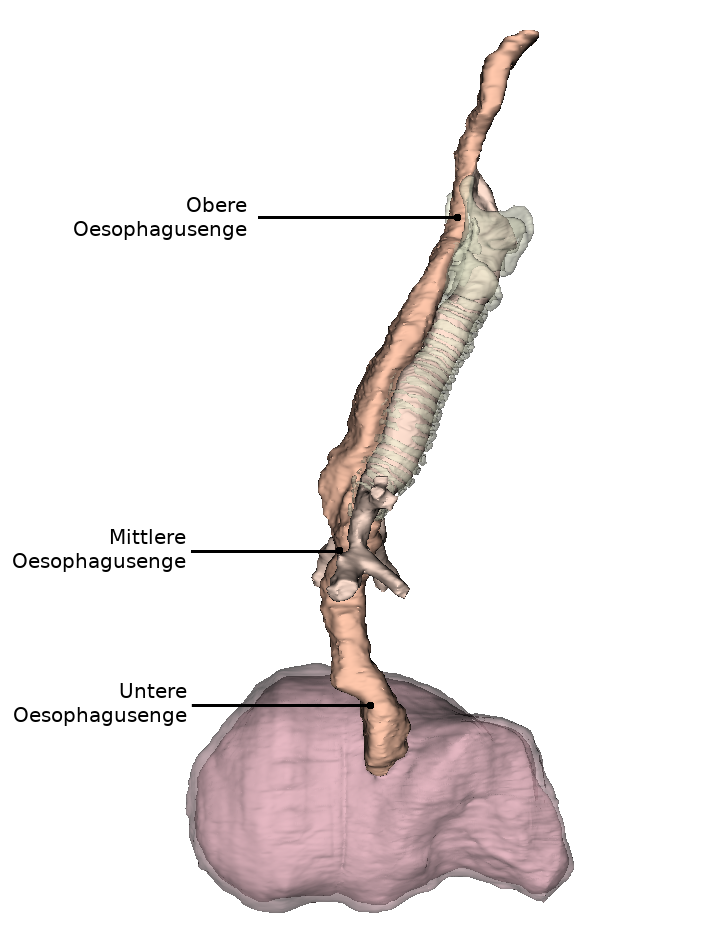
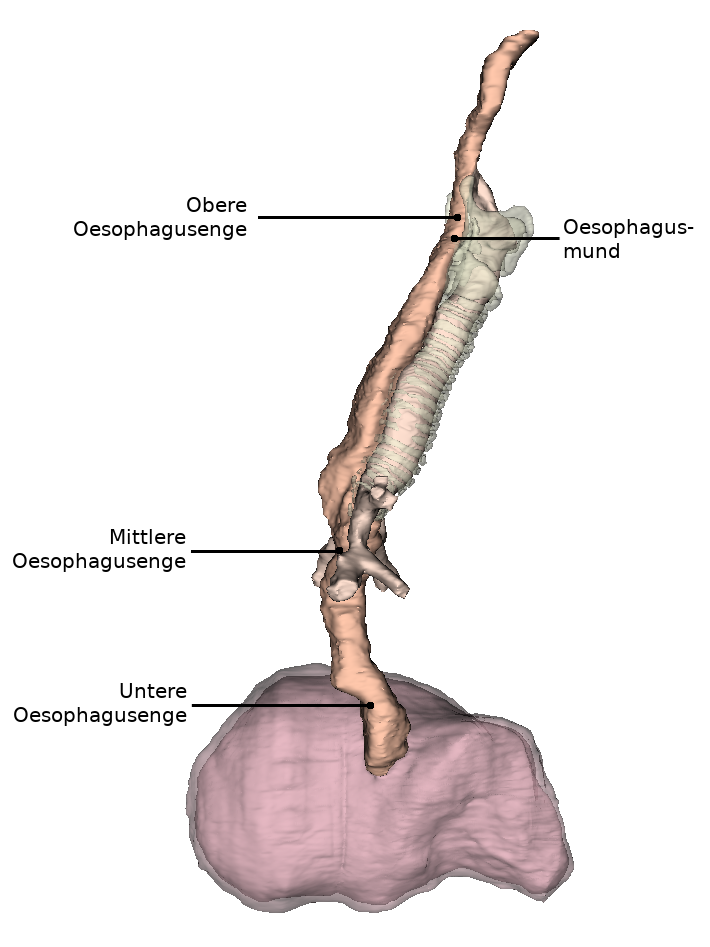
Due to its close relationship with neighboring organs, the esophagus has three narrowing points.
The esophageal orifice refers to the uppermost and narrowest part (constrictio pharyngooesophagealis). This is located in the pars cervicalis at the level of the cricoid cartilage of the larynx and measures approximately 14 millimeters in maximum diameter. Starting from the cricoid cartilage of the larynx, muscles surround the esophageal orifice and close it in the form of a sphincter, which is closed at rest.
The middle constriction (Constrictio partis thoracicae) in the pars thoracica is also called aortic stenosis. This is caused by the constriction of the esophagus by the left main bronchus and the aortic arch in the area of BWK 4/5. The maximum diameter here is 14 millimeters.
The inferior constriction (constrictio phrenica) results from the esophagus passing through the diaphragm at the beginning of the pars abdominalis and is called diaphragmatic constriction. Functional closure of the esophagus is ensured by the adjacent longitudinal tension of the musculature and by the veins of the esophageal wall. At rest (outside the act of swallowing), the lower part of the esophagus (pars abdominalis) is permanently closed.
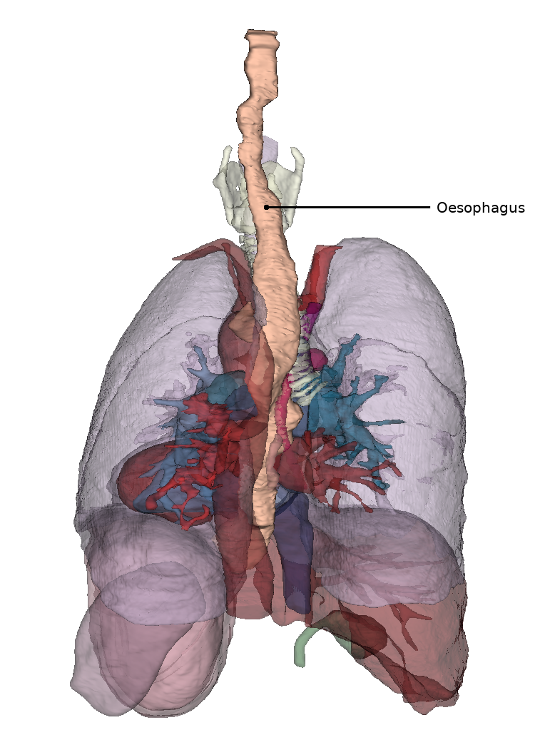
Lage im Horizontalschnitt
In der Sicht auf den Thorax von ventral liegt die Speiseröhre überwiegend im unteren Teil und ist bezogen auf die Medianlinie durch die links von ihr verlaufende Aorta leicht nach rechts verschoben. Ein Stück unterhalb des Proc. xiphoideus sterni tritt sie durch das Zwerchfell in die Peritonealhöhle ein.
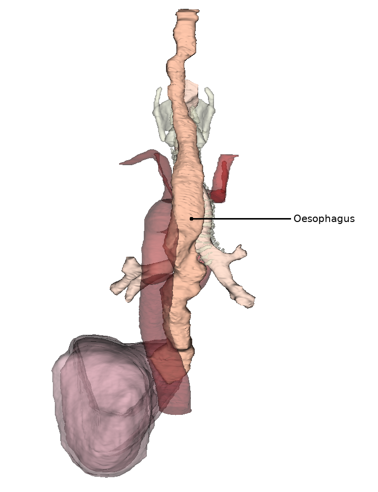
Beziehung zu Nachbarorganen
Unmittelbar an der vorderen Seite der Speiseröhre verläuft die Luftröhre bis zu ihrer Aufteilung in die beiden Hauptbronchien (lat. Bifurcatio tracheae). Durch eine Vielzahl von Bindegewebszügeln ist die Vorderwand der Speiseröhre mit der Rückwand der Luftröhre verbunden.
Unterhalb der Struktur der Luftröhrengabel, im unteren Mediastinum (lat. Mediastinum inferius), liegt die Speiseröhre eng am linken Herzvorhof und der Aorta thoracica an. Weiterhin verläuft die Aorta zunächst links der Speiseröhre nach kaudal und kommt kurz vor dem Eintritt der Speiseröhre in das Zwerchfell hinter dieser zu liegen.
Lage In situ
Die Speiseröhre weist neben den Engstellen auch charakteristische Krümmungen auf.
Die obere Krümmung verläuft in der Ansicht von ventral in der Pars cervicalis nach links. Die mittlere Krümmung liegt im Bereich der Pars thoracica und verläuft bedingt durch die nahe Aorta thoracica nach rechts. Die untere Krümmung in der Pars abdominalis wendet sich nun wieder nach links.
Betrachtet man die Speiseröhre in der Sagittalebene, so ist zu erkennen, dass diese dem Wirbelsäulenverlauf folgt und leicht konkav nach vorn gebogen ist.
Erkrankungen