Toe bones
The big toe (hallux) consists of two limbs (phalanx proximalis and distalis), the remaining toes of three (phalanx proximalis, media and distalis). Like the metatarsals, the proximal phalanges are divided into base, corpus and caput.
Anatomy


The toe bones are formed by 14 tubular bones. These represent the phalanges of the foot. They are the end phalanges of the foot and therefore connect distally to the five metatarsal bones. The individual bony elements are divided between the individual toes. All toes consist of three phalanges, except the big toe, which consists of only two phalanges. Due to this division, the phalanges are called differently. The toe elements close to the body are the phalanx proximalis. The distal phalanges are called phalanx distalis. Between these are the phalanx media. In big toes the phalanx media is missing, so it consists only of the phalanx proximalis and phalanx distalis.
The individual phalanges can also be structurally subdivided according to their affiliation to the tubular bones. A single one of these bone elements is subdivided into the head, the shaft and the base. The head is called caput phalangis and represents the distal end of the bone. It is flattened at the distal phalangis, where no other bone is adjacent.
Das proximale Ende des Röhrenknochens heißt Basis phalangis. Beim Phalanx proximalis hat dieser Teil eine konkave Grundfläche, die in Verbindung zum jeweiligen Mittelhandknochen steht.
Zwischen der Basis und dem Kopf der Phalangen befindet sich der Schaft, der Corpus phalangis. Er ist ein etwas schmalerer Bereich und hat eine säulen-ähnliche Form.
Das Phalanx proximalis wird auch Zehengrundglied genannt und stellt den längsten der jeweils drei Knochenglieder dar. Seine Basis trifft jeweils auf den zugehörigen Mittelfußknochen. Das distale Ende artikuliert bei den Zehen jeweils mit dem Phalanx media. Ausnahme davon ist wieder die Großzehe. Dort ist der Phalanx proximalis direkt mit dem Phalanx distalis verbunden.
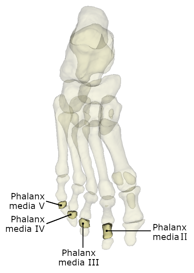
Das Zehenmittelglied (Phalanx media) Steht in der Mitte der Phalangen-Reihe. Es hat eine mittlere Größe und seine Basis hat einen größeren Durchmesser als das distale Ende. Am Kopf dieses Phalangen sind jeweils zwei kleine knöcherne Erhebungen zu finden. Das proximale Ende schließt sich an die Zehengrundglieder an. Am körperfernen Ende dagegen folgt das Phalanx distalis.
Das körperfernste Glied der gesamten oberen Extremität ist das Zehenendglied oder auch Phalanx distalis. Es ist jeweils der kürzeste der Zehenglieder. Die Basis artikuliert mit dem Phalanx media, außer an der Großzehe dort ist es direkt mit dem Zehengrundglied verknüpft.