Kneecap
The kneecap (patella) is a flat, disc-like bone. It is the largest sesamoid bone in the body, which means it is completely embedded in a tendon.
Anatomy
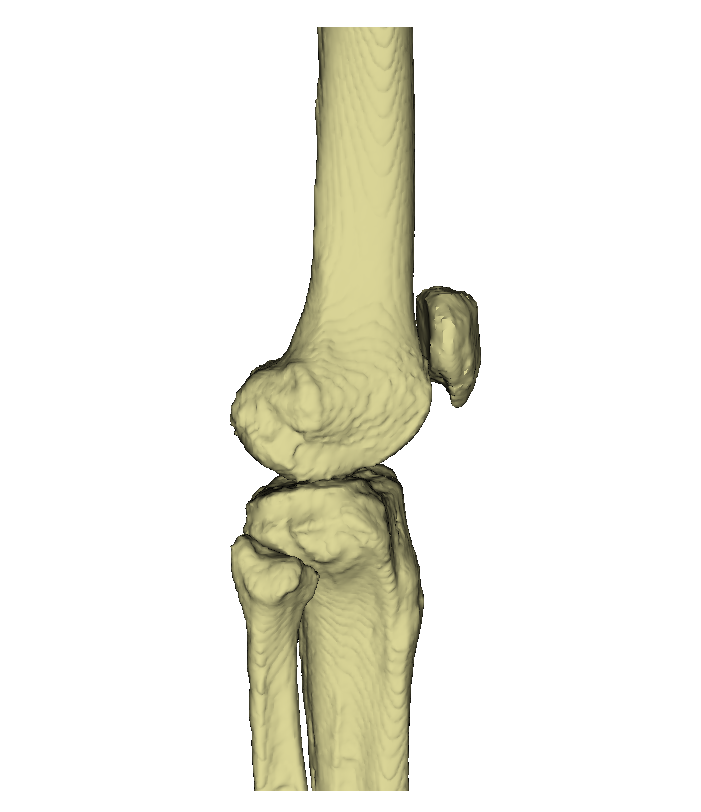


The kneecap is located in front of the knee joint. It is a flat bone with several edges and surfaces.
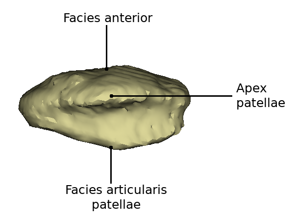
The front side is called facies anterior. It has a convex curvature and is completely covered by the tendon of the musculus quadriceps femoris. The facies posterior is divided into two halves by a depression (sulcus intercondylaris). The lateral side is wider than the medial side. The angle between the two halves varies, but is usually between 120° and 140°. Two thirds of the posterior surface is covered with articular cartilage.
The upper edge of the patella is very strong and is called Margo superior. The inner margin (Margo medialis) thins out towards the distal side. The outer edge is called Margo lateralis and also becomes thinner as it progresses. The lower tip, which is formed between the outer and inner margin is called apex patellae.
Function
The kneecap is used to transmit the force from the thigh. The kneecap increases the distance to the centre of rotation, resulting in a saving of force. The cartilage layer reduces the sliding resistance of the patella tendon. Furthermore, the patella takes over a protective function for the thigh bone. When stretching, the kneecap performs a movement of 8-10 cm above the thigh bone.