Magen (Gaster)/en: Unterschied zwischen den Versionen
Becher (Diskussion | Beiträge) (Die Seite wurde neu angelegt: „The largest part of the stomach is formed by the gastric body (<i>corpus gastricum</i>). The stomach ends at the top with the <i>fundus gastricus</i>, which is…“) |
Becher (Diskussion | Beiträge) |
||
| (40 dazwischenliegende Versionen von 2 Benutzern werden nicht angezeigt) | |||
| Zeile 1: | Zeile 1: | ||
| − | < | + | The stomach (lat. gaster) is a hollow-spaced digestive organ. In the stomach, gastric juice and digestive pulp are mixed together and uniformly tempered. |
| + | |||
| + | <div class="button_style"> | ||
| + | <div class ="center"> | ||
| + | <div class="dropdown"> | ||
| + | <div class="floatright" style="margin:0.4em;">[[File:PfeilDropdown.png|20px|link=|Exercises]]</div> | ||
| + | <div class="dropbtnart">[[Special:MyLanguage/Übungsaufgaben|Exercises]]</div> | ||
| + | <div class="dropdown-content"> | ||
| + | <div>[[Special:MyLanguage/Übungsaufgaben: Abdomen und Becken|Abdomen and pelvis]]</div> | ||
| + | <div>[[Special:MyLanguage/Übungsaufgabe: Magen|Stomach]]</div> | ||
| + | </div> | ||
| + | </div> | ||
| + | </div> | ||
| + | </div> | ||
| + | |||
| + | <div class="box"> | ||
| + | <div class="pic">__TOC__</div> | ||
| + | <div class="segmenter"><segmenter-embedding public wsemb-id="Magen" file="Magen.seg" height="300" width="400"/></div> | ||
| + | </div> | ||
| − | == | + | <div class="dropdown"> |
| + | <div class="floatright" style="margin:0.4em;">[[File:PfeilDropdown.png|20px|link=|Neigbouring structures]]</div> | ||
| + | <div class="dropbtnart">Neigbouring structures</div> | ||
| + | <div class="dropdown-content"> | ||
| + | <div>[[Special:MyLanguage/Speiseröhre|Esophagus]]</div> | ||
| + | <div>[[Special:MyLanguage/Milz|Spleen]]</div> | ||
| + | <div>[[Special:MyLanguage/Leber|Liver]]</div> | ||
| + | <div>[[Special:MyLanguage/Lunge|Lung]]</div> | ||
| + | </div> | ||
| + | </div> | ||
| − | <div style=" | + | <div class="dropdown"> |
| − | < | + | <div class="floatright" style="margin:0.4em;">[[File:PfeilDropdown.png|20px|link=|Body regions]]</div> |
| + | <div class="dropbtnart">[[Special:MyLanguage/Körperregionen|Body regions]]</div> | ||
| + | <div class="dropdown-content"> | ||
| + | <div>[[Special:MyLanguage/Organe Abdomen und Becken|Organs of abdomen and pelvis]]</div> | ||
| + | <div>[[Special:MyLanguage/Innere Organe|Inner organs]]</div> | ||
| + | <div>[[Special:MyLanguage/Rumpf|Trunk]]</div> | ||
| + | </div> | ||
</div> | </div> | ||
| − | + | <div class="dropdown"> | |
| − | + | <div class="floatright" style="margin:0.4em;">[[File:PfeilDropdown.png|20px|link=|Organ system]]</div> | |
| + | <div class="dropbtnart">[[Special:MyLanguage/Organsystem|Organ system]]</div> | ||
| + | <div class="dropdown-content"> | ||
| + | <div>[[Special:MyLanguage/Verdauungsapparat|Digestive system]]</div> | ||
| + | </div> | ||
| + | </div> | ||
| + | ---- | ||
| + | ==Anatomy== | ||
| + | The largest part of the stomach is formed by the gastric body (corpus gastricum). The stomach ends at the top with the fundus gastricus, which is highest in the standing patient and where the inhaled air is located (visible as a stomach bladder in the lower CT image). The fundus is at about the same level as the horizontal boundary of the liver. The esophagus ends in the pars cardiaca, which is located in the upper part of the stomach. On the outside, the stomach is covered with serous peritoneal, while the esophagus is surrounded by adventitious connective tissue. </br> | ||
| + | After the corpus follows the antrum pyloricum. The transition of the porter's channel into the small intestine is formed by the m. sphincter pylori. The m. sphincter pylori is caused by the particularly strong ring muscles of the stomach. This causes the visible constriction of the canalis pyloricus. | ||
| − | |||
| − | < | + | ==Position in horizontal section== |
| − | + | <div class="thumb tright thumbinner"> | |
| + | <div class="picture"> | ||
| + | <lightbox-embedding src="file:Magenschnitt.png" group="image-group-1" caption="Stomach: Section through the stomach. View from cranial with section in the horizontal in the region of the 1st lumbar or 7th thoracic vertebra." width="400" height="300" style="width:300px; height:300px;float:left;margin:1px;background-color:#fff;border:1px solid #c8ccd1;display: flex;justify-content: center;"/> | ||
| + | </div> | ||
| + | <div class="thumbcaption"> | ||
| + | Stomach: Section through the stomach</div> | ||
| + | </div> | ||
| + | The stomach lies within the peritoneum (intraperitoneal) and is located in the immediate vicinity of various organs. The curved organ contours on both sides of the stomach are called large (curvaturagastrica major) and small (curvaturagastrica minor) stomach curvatures. Ventrally, the left lobe of the liver towers above the stomach up to the left upper abdomen. The stomach is dorsally adjacent to the spleen. The stomach has two mesogastria as developmental rudiments, which develop further as omentum majus and minus as well as connecting lines between stomach and adjacent organs and include the net bag (bursa omentalis). The bursa omentalis is the slit-shaped displacement space of the peritoneal cavity, which lies dorsally from the stomach. The back wall of the bursa omentalis is formed by the pancreas. For gastric peristalsis, the peritoneal coating plays an important role, as it establishes mobility in relation to the neighbouring organs. | ||
| + | <br><br> | ||
| − | + | ==Projection onto the trunk== | |
| + | <div class="thumb tright thumbinner"> | ||
| + | <div class="picture"> | ||
| + | <lightbox-embedding src="file:gaster.png" group="image-group-1" caption="Stomach: Projection onto the trunk" width="400" height="300" style="width:300px; height:300px;float:left;margin:1px;background-color:#fff;border:1px solid #c8ccd1;display: flex;justify-content: center;"/> | ||
| + | </div> | ||
| + | <div class="thumbcaption"> | ||
| + | Stomach: Projection onto the trunk</div> | ||
| + | </div> | ||
| + | The gastric fundus/corpus is projected on the trunk between the 6th and 10th rib, on the area between linea mediana anterior and linea medioclavicularis. The path between the symphysis upper edge and the upper edge of the manubrium sterni plays an important role in clinical investigations. The pyloric part is located in this area (pylorus). The pyloric part is rather immovable from the rest of the stomach because he is connected to the duodenum located in the Retroperitoneum. | ||
<br> | <br> | ||
| − | |||
| − | |||
| + | ==Relationship to neighbouring organs== | ||
| − | + | Due to the position of the stomach within the peritoneum, it is very mobile in relation to neighbouring organs. Under certain circumstances, this can lead to disease processes (stomach ulcers, malignant tumours) that penetrate the stomach wall spreading to tight-fitting, neighbouring organs. | |
| + | <br> | ||
| − | + | ==Stomach in situ== | |
| − | + | Stomach in situ from the anterior view of the abdomen with elevated liver and removed omentum minus or major. In the left upper abdomen the close contact between the large gastric curvature and the spleen becomes visible. | |
| − | |||
| + | ==Diseases== | ||
| + | *[[Special:MyLanguage/Reizmagen|Irritable stomach]] | ||
| + | *[[Special:MyLanguage/Magenschleinhautentzündung|Gastritis]] | ||
| + | *[[Special:MyLanguage/Morbus Crohn|Morbus Crohn]] | ||
| + | *[[Special:MyLanguage/Colitis ulcerosa|Colitis ulcerosa]] | ||
| − | == | + | ==Free exploration== |
| + | <div style="float:left;margin-right:1em;"><segmenter-embedding public wsemb-id="Magen" file="Magen.seg" height="300" width="400"/></div> | ||
| − | + | <div style="float:left;width:50%">Look at the structure of the stomach in 3D and explore it freely. Afterwards you can test your acquired knowledge by the exercises.</div> | |
| − | < | + | <div class="clear"></div> |
<div class="clear"></div> | <div class="clear"></div> | ||
| − | <div style=" | + | ---- |
| − | + | <div class="clear aufgaben" style="margin-bottom:1em;"> | |
| − | + | <div class="menu_item"> | |
| − | < | + | <li class="mw-ui-button button_new" >[[Special:MyLanguage/Übungsaufgaben|Exercises]]</li> |
| + | </div> | ||
</div> | </div> | ||
| − | |||
| + | <div class="clear aufgaben"> | ||
| + | <div class="menu_item"> | ||
| + | <li class="button_article"><b>Further article</b></li> | ||
| + | </div> | ||
| − | = | + | <div class="menu_item"> |
| − | + | <li class="mw-ui-button button_normal">[[Special:MyLanguage/Herz|Heart]]</li> | |
| − | + | </div> | |
| − | |||
| + | <div class="menu_item"> | ||
| + | <li class="mw-ui-button button_normal">[[Special:MyLanguage/Gallenblase|Gallbladder]]</li> | ||
| + | </div> | ||
| + | </div> | ||
| + | <div class="clear"></div> | ||
---- | ---- | ||
| + | [[Category:Organs abdomen]] | ||
| + | [[Category:Inner organs]] | ||
| + | [[Category:Organ system]] | ||
| + | [[Category:Body regions]] | ||
| − | + | </div> | |
| − | + | <languages/> | |
| − | |||
| − | |||
| − | |||
Aktuelle Version vom 4. Januar 2022, 14:54 Uhr
The stomach (lat. gaster) is a hollow-spaced digestive organ. In the stomach, gastric juice and digestive pulp are mixed together and uniformly tempered.
Anatomy
The largest part of the stomach is formed by the gastric body (corpus gastricum). The stomach ends at the top with the fundus gastricus, which is highest in the standing patient and where the inhaled air is located (visible as a stomach bladder in the lower CT image). The fundus is at about the same level as the horizontal boundary of the liver. The esophagus ends in the pars cardiaca, which is located in the upper part of the stomach. On the outside, the stomach is covered with serous peritoneal, while the esophagus is surrounded by adventitious connective tissue.
After the corpus follows the antrum pyloricum. The transition of the porter's channel into the small intestine is formed by the m. sphincter pylori. The m. sphincter pylori is caused by the particularly strong ring muscles of the stomach. This causes the visible constriction of the canalis pyloricus.
Position in horizontal section
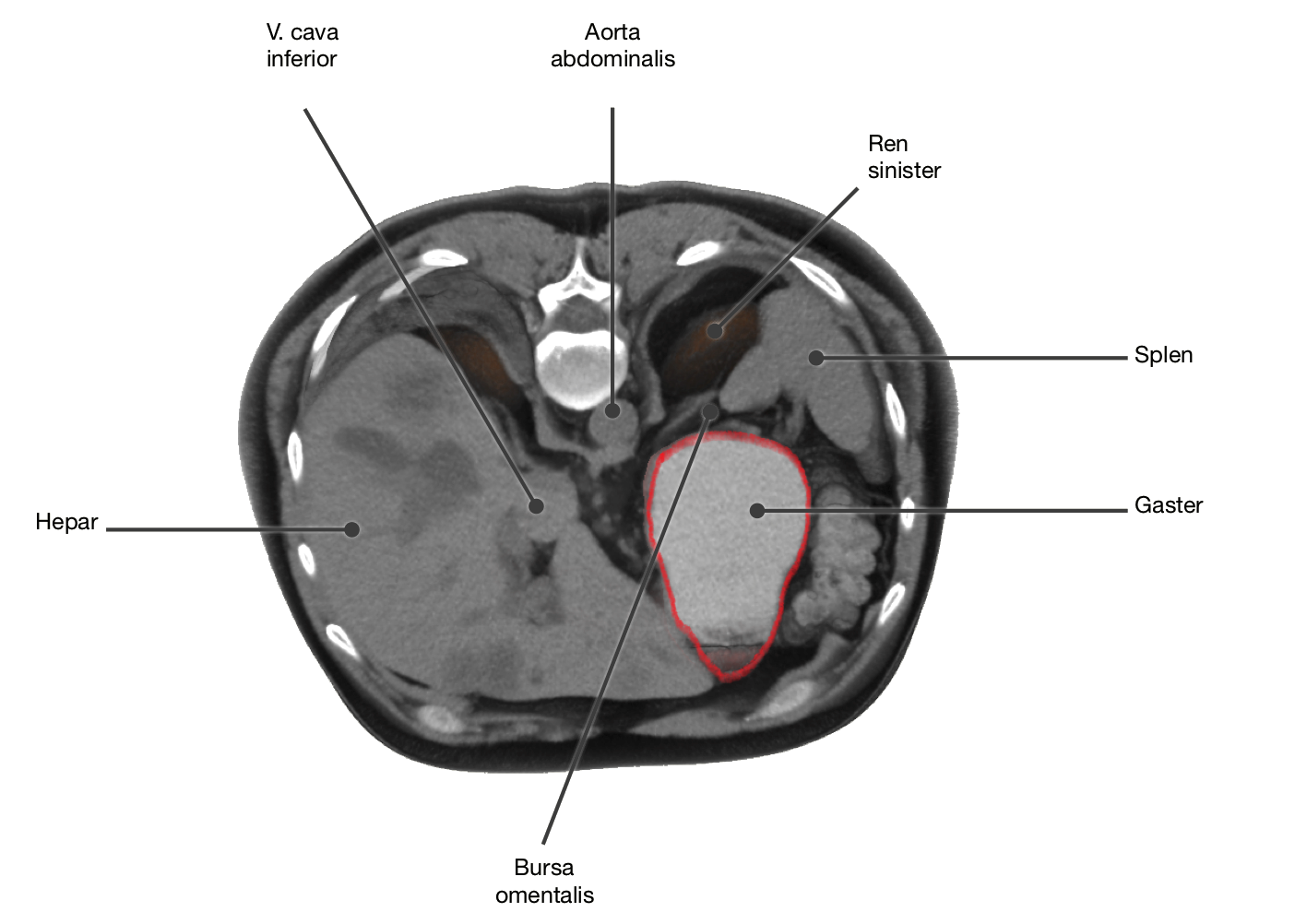
The stomach lies within the peritoneum (intraperitoneal) and is located in the immediate vicinity of various organs. The curved organ contours on both sides of the stomach are called large (curvaturagastrica major) and small (curvaturagastrica minor) stomach curvatures. Ventrally, the left lobe of the liver towers above the stomach up to the left upper abdomen. The stomach is dorsally adjacent to the spleen. The stomach has two mesogastria as developmental rudiments, which develop further as omentum majus and minus as well as connecting lines between stomach and adjacent organs and include the net bag (bursa omentalis). The bursa omentalis is the slit-shaped displacement space of the peritoneal cavity, which lies dorsally from the stomach. The back wall of the bursa omentalis is formed by the pancreas. For gastric peristalsis, the peritoneal coating plays an important role, as it establishes mobility in relation to the neighbouring organs.
Projection onto the trunk
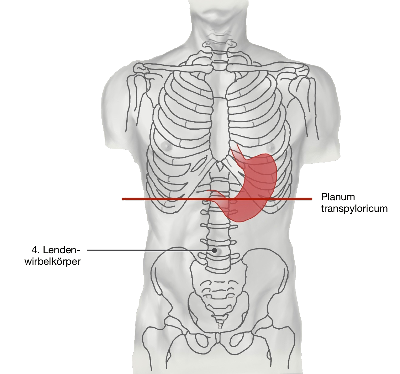
The gastric fundus/corpus is projected on the trunk between the 6th and 10th rib, on the area between linea mediana anterior and linea medioclavicularis. The path between the symphysis upper edge and the upper edge of the manubrium sterni plays an important role in clinical investigations. The pyloric part is located in this area (pylorus). The pyloric part is rather immovable from the rest of the stomach because he is connected to the duodenum located in the Retroperitoneum.
Relationship to neighbouring organs
Due to the position of the stomach within the peritoneum, it is very mobile in relation to neighbouring organs. Under certain circumstances, this can lead to disease processes (stomach ulcers, malignant tumours) that penetrate the stomach wall spreading to tight-fitting, neighbouring organs.
Stomach in situ
Stomach in situ from the anterior view of the abdomen with elevated liver and removed omentum minus or major. In the left upper abdomen the close contact between the large gastric curvature and the spleen becomes visible.