Herz/en: Unterschied zwischen den Versionen
Becher (Diskussion | Beiträge) (Die Seite wurde neu angelegt: „Category:Organsystem/en“) |
Will (Diskussion | Beiträge) |
||
| Zeile 60: | Zeile 60: | ||
<!--[segmenter_snapshot aufbau 3][/]--><b>View from dorsal</b> | <!--[segmenter_snapshot aufbau 3][/]--><b>View from dorsal</b> | ||
| − | From <!--<segmenter-snapshot wsemb_id="aufbau" snapshot_index="6">-->this perspective <!--</segmenter-snapshot>--> it is clearly visible how the Arcus aortae crosses the Truncus pulmonalis, where it divides into the A. pulmonalis sinistra and A. pulmonalis dextra. At this point the three major arteries branch off to the upper extremity and to the neck and skull: Truncus brachiocephalicus, A. carotis communis | + | From <!--<segmenter-snapshot wsemb_id="aufbau" snapshot_index="6">-->this perspective <!--</segmenter-snapshot>--> it is clearly visible how the Arcus aortae crosses the Truncus pulmonalis, where it divides into the A. pulmonalis sinistra and A. pulmonalis dextra. At this point the three major arteries branch off to the upper extremity and to the neck and skull: Truncus brachiocephalicus, A. carotis communis sinistra and A. subclavia sinistra. Also the orifices of the - usually four - Vv. pulmonales into the left atrium (Atrium sinistrum) and the two Vv. cavae into the right atrium (Atrium dextrum) are well visible. Also visible here is the sinus coronarius in the sulcus of the same name. This sinus is the collection vessel for the venous blood that is supplied to the heart via the Vv. cardiacae. |
<br> | <br> | ||
<div class="clear"> | <div class="clear"> | ||
Version vom 9. November 2020, 08:35 Uhr
The heart (lat. Cor) is a hollow muscle. Through its contractions the blood is pumped through the body. It is a vital organ that ensures the necessary supply of blood to the other organs.
Heart in situ
The heart is located approximately in the middle of the thorax, which is called the mediastinum. The representation in the WebViewer is greatly simplified.
In order to have a clear view of the heart, the thorax must be opened extensively and the connective tissue in the mediastinum anterius removed. Approximately two-thirds of the heart is located to the left of the breastbone and one-third to the right. The apex of the heart (apex cortis) points to the left front and lies on one level with the left nipple. Its contraction (the so-called cardiac apex thrust) can be felt as a tender beat through the chest wall. Viewed ventrally, the heart is located obliquely and turned counterclockwise in the thorax. At the front it reaches the sternum and at the back it is bounded by the trachea and oesophagus. On the left and right it is surrounded by the lungs.
The right ventricle is clearly visible from the ventral view. The left ventricle is only partially visible.
Even the large vessels are not all visible at the base of the heart. The Vv. pulmonales lies on the flip side of the heart and merges in the left atrium, which is also dorsal. Clearly recognizable, however, are the two heart ears (Auricula sinistra and dextra), each located on the periphery. The epicardium surrounding the heart is not visible in this representation.
Form and structure
View from ventral
The heart (Cor) has the shape of an obliquely inclined cone and is a muscular hollow organ.
The following structures are differentiated in the heart:
- a downward, left and ventrally inclined tip of the heart,
- three areas, whose names are mostly based on the adjacent thorax walls
- a heart base inclined upwards, to the right and dorsally
On the side of the heart that lies on the sternum and costae, the right ventricle is visible, separated from the left ventricle by the anterior interventricular sulcus. From this angle the left ventricle forms the left edge of the heart and the apex cordis.
The anterior intercostal furrow (sulcus interventricularis anterior) contains the R. interventricularis anterior, the A. coronaria sinistra and the V. interventricularis anterior. The sulcus interventricularis anterior describes the course of the ventricular septum inside the heart.
The atria (atrium sinistrum and dextrum)are separated from the ventricles by the sulcus coronarius which also contains coronary vessels.
The right auricle (Auricula dextra) attaches to the base of the aorta (Pars ascendens), the left auricle (Auricula sinistra) to the base of the truncus pulmonalis. From this perspective the aorta hides the exit of the right pulmonary artery from the pulmonary trunk.
View from dorsal and caudal
By turning the heart ventrally, the side facing the diaphragm (Fascies diaphragmatica) becomes more visible. Exclusively from caudally from the viewing direction of the diaphragm it can be seen that the both Vv. cavae lie in one axis.
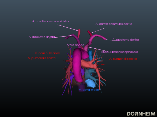
View from dorsal
From this perspective it is clearly visible how the Arcus aortae crosses the Truncus pulmonalis, where it divides into the A. pulmonalis sinistra and A. pulmonalis dextra. At this point the three major arteries branch off to the upper extremity and to the neck and skull: Truncus brachiocephalicus, A. carotis communis sinistra and A. subclavia sinistra. Also the orifices of the - usually four - Vv. pulmonales into the left atrium (Atrium sinistrum) and the two Vv. cavae into the right atrium (Atrium dextrum) are well visible. Also visible here is the sinus coronarius in the sulcus of the same name. This sinus is the collection vessel for the venous blood that is supplied to the heart via the Vv. cardiacae.
Inner spaces of the heart
In the heart, four internal spaces are distinguished (one chamber (ventricle) and one atrium (atrium) on the left and right respectively). The atria are adjacent to the ventricles, which are connected to the lungs or the major arteries and veins of the body by their outgoing vessels.
Inner spaces of the left heart
The left atrium has smooth walls between the openings of the four pulmonary veins (Vv. pulmonales dextra/sinistra superior and inferior). The muscle wall of the left atrium is thinner than that of the right atrium because it belongs to the low pressure system. The narrow fold (valvula foraminis ovalis) is temporarily visible at the septum interatriale. It is formed by raising the fossa ovalis into the left atrium.
Blood enters the left ventricle via the ostium atrioventriculare sinistrum from the left atrium.. The atrioventricular ostium can be closed by the sinistra atrioventricular valve. In addition the left ventricle has an inflow and outflow tract. Whereby the inflow runs along the posterior wall, the left lateral wall as well as the apical section. The smooth-walled outflow tract is located near the septum interventriculare and continues into the vestibulum aortae. It consists mainly of muscles (pars muscularis). The ostium aortae is the opening between the aortic outflow tract of the left ventricle and the aorta. The wall of the left ventricle is about three times as thick as that of the right ventricle and is therefore part of the high pressure system.
Inner spaces of the right heart
The posterior part of the right atrium consists of the sinus venarum cavarum (atrial sinus). The two Vv. cavae (superior and inferior) join at the ostium cavea superioris and inferioris. From these the blood can flow seamlessly into the atrium without intermediate valves. The fossa ovalis is located above the confluence of the V. cava inferior and is bordered by the limbus fosssae ovalis. The crista terminalis separates the anterior part (right atrium with a heart-ear) from the posterior part.
The right atrium is larger than the left and is also part of the low pressure system. In contrast to the smooth-walled front part of the atrium, the rear part has a much more complex structure.
Through the trabeculae carnea and the crista supraventricularis the right ventricle can also be divided into two parts.
The blood flows via the ostium atrioventriculare dextrum into the right ventricle. Here, too, you can see a inflow and outflow path. In the inflow area there are the Trabeculae carneae, which are small muscle clusters. Also here the Mm. papillares are connected via Chordae tendineae with the Valva atrioventricularis (dextra). The outflow tract continues above the conus arteriosus. The conus arteriosus is the conical transition of the right ventricle into the truncus pulmonalis. This area is smooth-walled and does not show any Trabeculae carneae. The blood flows via the pulmonary valve (valva trunci pulmonalis) through the ostium trunci pulmonalis into the truncus pulmonalis. The right ventricle also belongs to the low pressure system.
Overview of the heart valves(Coming soon)
There are two types of valves in the heart: atrial-ventricular valves and vascular valves.
Cardiac auscultation (Coming soon)
In the case of a functional disorder, audible flow noises are produced by vortex formations of the blood.
Excitation formation and excitation conduction system (Coming soon)
In the heart there is an autonomous excitation system (Systema conducente cordis) and an excitation conduction system (Systema conducente cordis). Four sections are distinguished.
Sinus node
also Nodus sinuatrialis or Keith-Flack node
Atrioventricular node
also Nodus atrioventricularis, AV node or Aschoff-Tawara node
Atrioventricular bundle
also Fasciculus atrioventricularis, AV bundle or His bundle
Bundle branch
also Crus dextrum and sinistrum or Tawara-Schenkel
Diseases
Free exploration