The heart (lat. Cor) is a hollow muscle. Through its contractions the blood is pumped through the body. It is a vital organ that ensures the necessary supply of blood to the other organs.
Heart in situ
The heart is located approximately in the middle of the thorax, which is called the mediastinum. The representation in the WebViewer is greatly simplified.
In order to have a clear view of the heart, the thorax must be opened extensively and the connective tissue in the mediastinum anterius removed. Approximately two-thirds of the heart is located to the left of the breastbone and one-third to the right. The apex of the heart (apex cortis) points to the left front and lies on one level with the left nipple. Its contraction (the so-called cardiac apex thrust) can be felt as a tender beat through the chest wall. Viewed ventrally, the heart is located obliquely and turned counterclockwise in the thorax. At the front it reaches the sternum and at the back it is bounded by the trachea and oesophagus. On the left and right it is surrounded by the lungs.
The right ventricle is clearly visible from the ventral view. The left ventricle is only partially visible.
Even the large vessels are not all visible at the base of the heart. The Vv. pulmonales lies on the flip side of the heart and merges in the left atrium, which is also dorsal. Clearly recognizable, however, are the two heart ears (Auricula sinistra and dextra), each located on the periphery. The epicardium surrounding the heart is not visible in this representation.
Form and structure
View from ventral
The heart (Cor) has the shape of an obliquely inclined cone and is a muscular hollow organ.
The following structures are differentiated in the heart:
- a downward, left and ventrally inclined tip of the heart,
- three areas, whose names are mostly based on the adjacent thorax walls
- a heart base inclined upwards, to the right and dorsally
On the side of the heart that lies on the sternum and costae, the right ventricle is visible, separated from the left ventricle by the anterior interventricular sulcus. From this angle the left ventricle forms the left edge of the heart and the apex cordis.
The anterior intercostal furrow (sulcus interventricularis anterior) contains the R. interventricularis anterior, the A. coronaria sinistra and the V. interventricularis anterior. The sulcus interventricularis anterior describes the course of the ventricular septum inside the heart.
The atria (atrium sinistrum and dextrum) are separated from the ventricles by the sulcus coronarius which also contains coronary vessels.
The right auricle (Auricula dextra) attaches to the base of the aorta (Pars ascendens), the left auricle (Auricula sinistra) to the base of the truncus pulmonalis. From this perspective the aorta hides the exit of the right pulmonary artery from the pulmonary trunk.
<b>View from dorsal and caudal</b>
By turning the heart ventrally, the side facing the diaphragm (Fascies diaphragmatica) becomes more visible. Exclusively from caudally from the viewing direction of the diaphragm it can be seen that the both Vv. cavae lie in one axis.
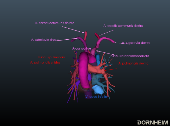
Ansicht von dorsal
Aus dieser Perspektive ist gut erkennbar, wie der Arcus aortae den Truncus pulmonalis überquert, wo sich dieser in die A. pulmonalis sinistra bzw. A. pulmonalis dextra aufteilt. An dieser Stelle zweigen die drei großen Arterien zur oberen Extremität und zu Hals und Schädel ab: Truncus brachiocephalicus, A.carotis communis sinisra und A. subclavia sinistra. Auch die Mündungen der - meist vier - Vv. pulmonales in den linken Vorhof (Atrium sinistrum) sowie der zwei Vv.cavae in den rechten Vorhof (Atrium dextrum) sind gut zu sehen. Ebenfalls sichtbar wird hier der Sinus coronarius im gleichnamigen Sulcus. Dieser Sinus ist das Sammelgefäß für das venöse Blut, das dem Herzen über die Vv.cardiacae zugeführt wird.
Binnenräume
Im Herzen werden vier Binnenräumen (jeweils links und rechts eine Kammer (Ventrikel) und ein Vorhof (Atrium)) unterschieden. Die Vorhöfe grenzen jeweils an die Ventrikel, die über ihre abgehenden Gefäße mit der Lunge oder den großen Körperarterien und -venen verbunden sind.
Binnenräume des linken Herzens
Der linke Vorhof ist glattwandig zwischen den Einmündungen der vier Lungenvenen (Vv. pulmonales dextra/sinistra superior und inferior). Die Muskelwand des linken Vorhofs ist dünner als die des rechten, da er zum Niederdrucksystem gehört. Die schmale Falte (Valvula foraminis ovalis) ist zeitweilig am Septum interatriale zu sehen. Sie entsteht durch emporragen der Fossa ovalis in den linken Vorhof.
Das Blut gelangt über das Ostium atrioventriculare sinistrum vom linken Vorhof in den linken Ventrikel. Das Ostium atrioventriculare kann dabei durch das Valva atrioventricularis sinistra verschlossen werden. Außerdem besitzt der linke Ventrikel eine Ein- und Ausstrombahn. Wobei der Einstrom entlang der Hinterwand, der linken Seitenwand wie auch des apikalen Abschnittes verläuft. Die glattwandige Ausstrombahn befindet sich in der Nähe vom Septum interventriculare und setzt sich in das Vestibulum aortae fort. Sie besteht größtenteils aus Muskulatur (Pars muscularis). Als Ostium aortae bezeichnet man die Öffnung zwischen der aortalen Ausstrombahn des linken Ventrikels und der Aorta. Die Wand des linken Ventrikels ist ca. drei mal so dick, wie die des rechten Ventrikels und gehört somit zum Hochdrucksystem.
Binnenräume des rechten Herzens
Der hintere Teil des rechten Vorhofs besteht aus dem Sinus venarum cavarum (Vorhofsinus). Am Ostium cavea superioris und inferioris münden die beiden Vv. cavae (superior und inferior). Von diesen kann das Blut nahtlos ohne dazwischenliegende Klappen in den Vorhof einfließen. Die Fossa ovalis befindet sich über dem Einmündungsort der V. cava inferior und wird vom Limbus fosssae ovalis umrandet. Die Crista terminalis grenzt den vorderen Teil (rechter Vorhof mit Herzohr) vom hinteren ab.
Der rechte Vorhof ist größer als der linke und gehört ebenfalls zum Niederdrucksystem. Im Gegensatz zum glattwandigen vorderen Abschnitts des Vorhofs, weißt der hintere Teil deutlich mehr Struktur auf.
Durch die Trabeculae carnea und die Crista supraventricularis lässt sich der rechte Ventrikel auch in zwei Teile trennen.
Das Blut fließt über das Ostium atrioventriculare dextrum in die rechte Herzkammer. Auch hier erkennt man eine Ein- und Ausflussbahn. Im Einstrombereich befinden sich die Trabeculae carneae, dabei handelt es sich um kleine Muskelbälkchen. Auch hier sind die Mm. papillares über Chordae tendineae mit der Valva atrioventricularis (dextra) verbunden. Die Ausflussbahn setzt über dem Conus arteriosus fort. Als Conus arteriosus bezeichnet man den kegelförmigen Übergang der rechten Herzkammer in den Truncus pulmonalis. Dieser Bereich ist glattwandig und weist keine Trabeculae carneae auf. Das Blut fließt über die Pulmonalklappe (Valva trunci pulmonalis) durch das Ostium trunci pulmonalis in den Truncus pulmonalis. Auch der rechte Ventrikel gehört zum Niederdrucksystem.
Übersicht über die Herzklappen (Demnächst)
Es werden zwei Arten von Klappen im Herzen unterschieden: Vorhof-Kammer-Klappen und Gefäßklappen.
Herzauskultation (Demnächst)
Bei einer Funktionsstörung entstehen durch Wirbelbildungen des Blutes hörbare Strömungsgeräusche.
Erregungsbildungs- und Erregungsleitungssystem (Demnächst)
Im Herzen gibt es ein autonomes Erregungsbildungssystem (Systema conducente cordis) und ein Erregungsleitungssystem (Systema conducente cordis). Es werden dabei vier Abschnitte unterschieden.
Sinusknoten
auch Nodus sinuatrialis oder Keith-Flack-Knoten
Atrioventrikularknoten
auch Nodus atrioventricularis, AV-Knoten oder Aschoff-Tawara-Knoten
Atrioventrikularbündel
auch Fasciculus atrioventricularis, AV-Bündel oder His-Bündel
Kammerschenkel
auch Crus dextrum und sinistrum oder Tawara-Schenkel
Erkrankungen
Freies Explorieren