Ribs
The human body has twelve pairs of ribs (lat. Costae). They have a curved and rod-like shape. The ribs run bent from the thoracic vertebrae to the sternum and form a cross connection. The connection between sternum and ribs is formed by the rib cartilage. The ribs represent an important part of the bony thorax.
Anatomy
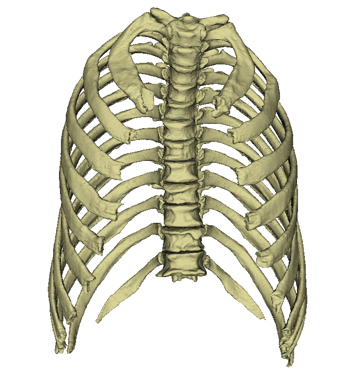
Ribs are the paired, curved, rod-shaped bones that originate on the dorsal side of the thoracic spine.
Most ribs then reach the sternum in a semicircle.
In this way, they create a cross connection between the two structures and thus serve the stability and elasticity of the thorax.
Usually the female chest is narrower and shorter than the male.
The human body has a total of twelve pairs of ribs, which corresponds to the number of thoracic vertebrae.
Each rib consists of the actual rib bone (Os costale) and an adjacent section of cartilage, the costal cartilage (Cartilago costalis). The individual ribs are not directly adjacent to each other, but are separated by the intercostal space. This space is partially filled by the intercostal muscles.
The ribs are connected to the spinal column by the rib heads. Most ribs touch two consecutive vertebrae. The joint surface of the head of the rib (Facies articularis capitis costae) is therefore divided in two in this case.
At the neck of the rib, the rib hump (tuberculum costae) is located, which is connected to the transverse process of the thoracic vertebra at the same level via a small joint surface (facies articularis tuberculi costae). Not far from the rib neck is the angulus costae (rib angle). The flat body of the rib has a convex outer surface (Facies externa) and a concave inner surface (Facies interna).
The anterior part of the ribs consists of cartilage. This elastically connects the ribs to the sternum. Without this freedom of movement, the ribs could not move when breathing in and out.
Classification of the ribs

The ribs can be divided into three groups. The decisive factor here is the connection to the sternum.
Die ersten sieben Rippenpaare erreichen als sogenannte echte Rippen (Costae verae) das Brustbein direkt über ihren zugehörigen Rippenknorpel.
The next five ribs are called false rib pairs (costae spuriae). The 8th, 9th and 10th ribs are only indirectly connected to the sternum. The respective nearest rib cartilages connect and are thus involved in the construction of the costal arch (arcus costalis).
The last two " false" pairs of ribs (costae fluctuantes) of the 11th and 12th ribs usually end up freely between the muscles of the lateral abdominal wall.
Unterschiedliche Rippenformen


Jedes Rippenpaar ist bilateral symmetrisch, die Form jedoch ist in jedem Segment verschieden. Der Rippenhals reicht vom Rippenköpfchen (Caput costae) bis zum Rippenhöckerchen (Tuberculum costae) und liegt am dorsalen Ende der Rippe. Er weist mit Ausnahme der 1. Rippe eine nach oben gerichtete Leiste (Crista colli costa) auf. Lateral vom Rippenhöckerchen biegt sich der Rippenkörper (Corpus costae) unter Bildung des Rippenwinkels (Angulus costae) nach vorne um. Die Rippenkörper der 2.-11. Rippe weisen unregelmäßige Krümmungen auf, die sogenannten Flächen- und Kantenkrümmungen.
Durch ihre zusätzliche Verdrehung um die Längsachse sind die Außenflächen der Rippen an ihrem vertebralen Ende etwas nach kaudal, und an ihrem ventralen Ende leicht nach kranial geneigt. Normalerweise sind die 1. und 12. Rippe am kürzesten, und die 7. Rippe am längsten. Der Rippenknorpel wiederrum nimmt von der 1.-7. Rippe an Länge zu, ab der 8. Rippe wird er wieder kürzer. Mit Ausnahme der 1., 11. und 12. Rippe besitzt jede Rippe an ihrer Unterfläche eine Furche (Sulcus costae), in dem die Interkostalgefäße und -nerven relativ geschützt verlaufen.
Funktion
Die Rippen bilden im Zusammenhang mit den Brustwirbeln, dem Rippenknorpel und dem Sternum den knöchernen Brustkorb. Ihre Hauptaufgabe liegt in der Stabilisierung und dem Schutz der Inneren Organe, vor allem von Herz und Lunge.
Bewegung
Die Rippen führen bei der Atmung eine maßgebliche Bewegung aus. Diese werden durch die Rippen-Wirbel-Gelenke und Rippen-Brustbein-Gelenke durchgeführt. Das Heben und Senken während des Ein- und Ausatmen wird durch die Intercostalmuskeln bedingt. Die Bewegung umfasst einen Rahmen von ca. 2 cm.
Entwicklung
Die Rippen entstehen aus Somiten. Die Knochenspangen verknöchern gegen Ende des zweiten Embryonalmonats. Dabei liegt der Ausgang der Verknöcherung in einigen Ossifikationskernen beim Angulus costae. Die Knochenspangen verfestigen sich nicht komplett, so haben auch erwachsene Menschen einen Rippenknorpel.