Liver (Hepar)
The liver is the largest digestive gland in the human body. Due to its function and tasks it is the central organ of metabolism. It forms important proteins and detoxifies the body. Almost all nutrients that are absorbed through the intestines are first passed through the liver.
Anatomy
Projection
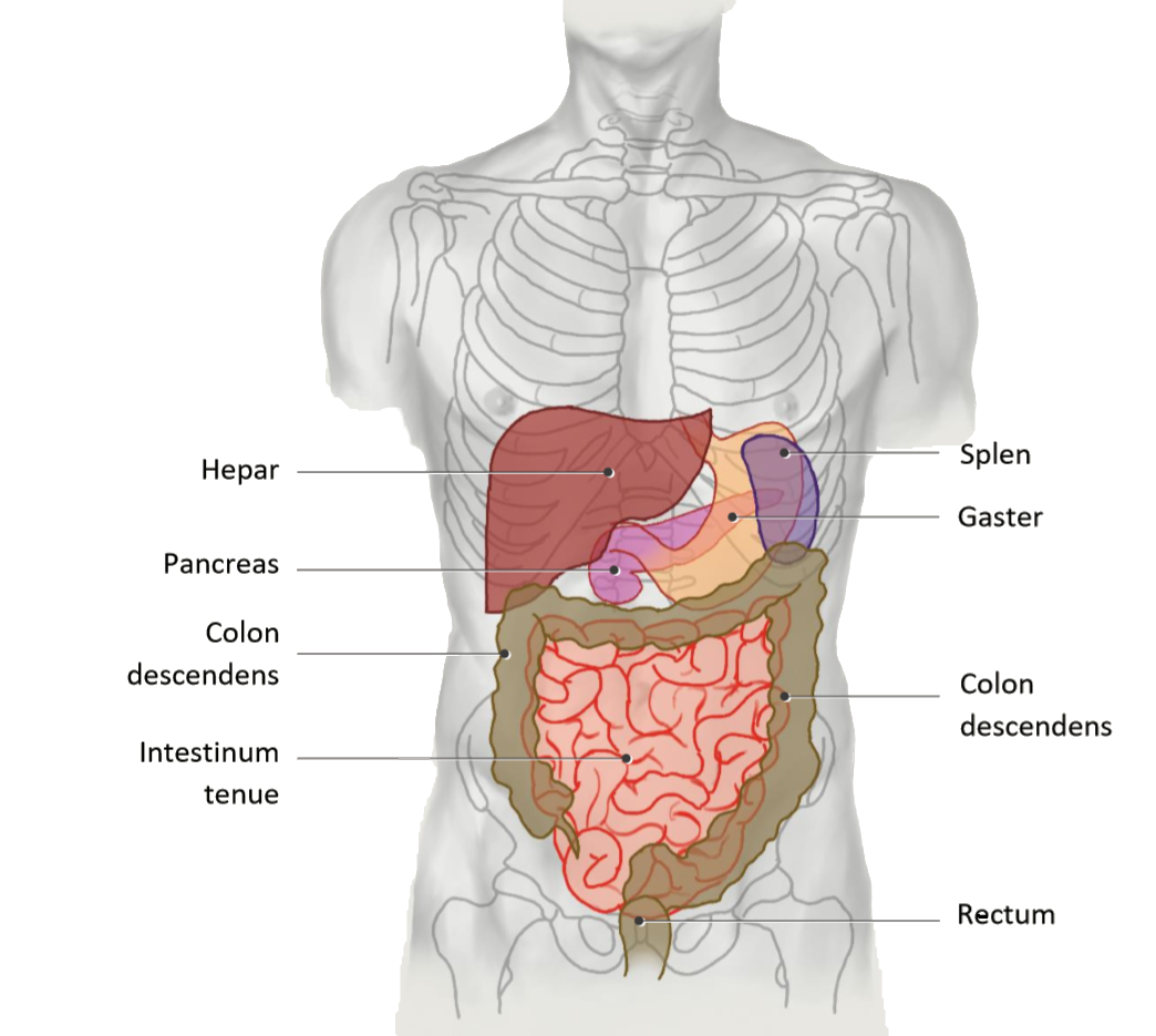
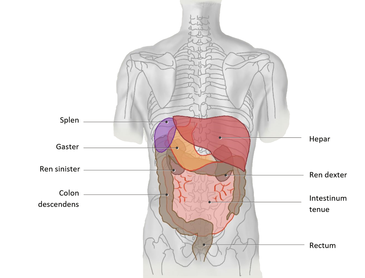
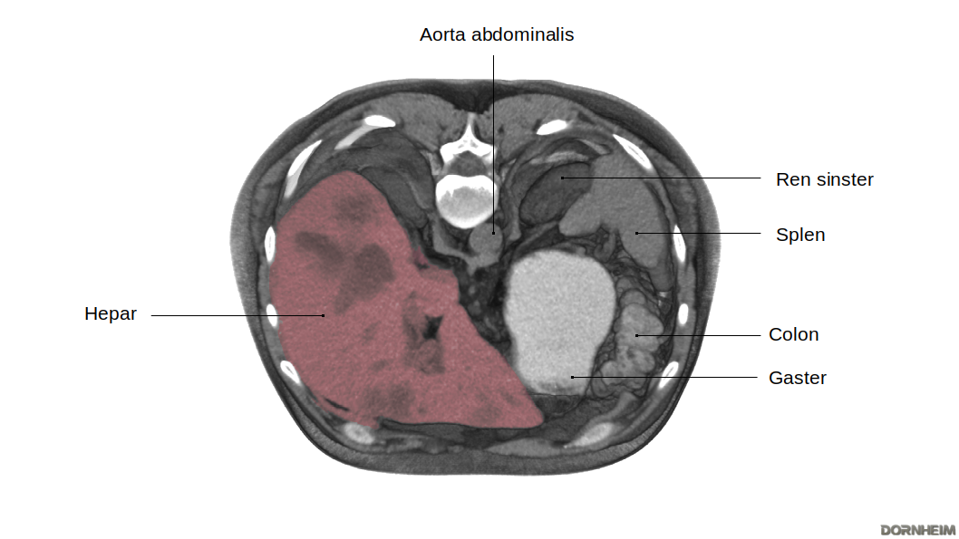
The liver is located in the right upper abdomen, but extends beyond the epigastrium into the left upper abdomen. It thereby pushes far in front of the stomach, which leaves an impression (impressio gastrica) at the back of the left liver lobe. The right lobe of the liver comes into close contact with the colon (right flexure), the small intestine (pars superior), the upper part of the right kidney and the right adrenal gland. Its impression sites are also named accordingly: Impressio colica, duodenalis, renalis and suprarenalis. The position of the liver is strongly dependent on respiration, as its underside is fused to the diaphragm. Age and posture are equally crucial in determining the position of the liver. The gallbladder is located dorsally between the left and right lobes of the liver.
Liver in situ
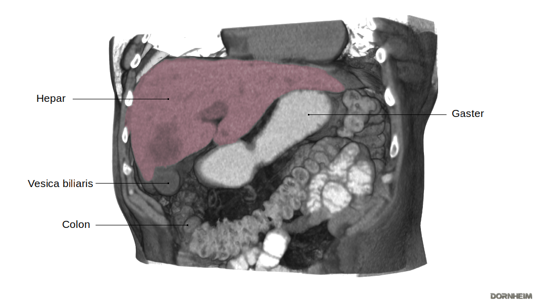
The thin area of tissue between the liver and the curvature of the stomach is called the omentum minus. This serous skin lines the peritoneal cavity and is the ventral border of the bursa omentalis. This is bordered on the right by the liver.
The downward-facing liver margin is sharp-edged and easily palpable in an enlarged liver. The liver extends from the right regio hypochondriaca, through the regio epigastrica, and into the left upper abdomen. The stomach can be seen at the left inferior border of the liver. The gallbladder lies on the underside of the liver and overhangs the inferior border of the liver.
Liver segments
The liver segments are formed by a total of 8 functional subunits of the liver. Each of these units has a branch of the portal vein, the bile duct and a segment branch of the arteria hepatica propria.
Assignment to Partes and Divisiones
| Pars hepatis sinistra | |
|---|---|
| Segment I | Pars posterior hepatis, Lobus caudatus |
|
Divisio lateralis sinistra |
| Segmentum mediale sinistrum (= Segmentum IV) subdivided into segment IVa and IVb |
Divisio medialis sinistra |
| Pars hepatis dextra | |
|---|---|
| Segmentum anterius mediale dextrum (= Segmentum V) Segmentum posterius mediale dextrum |
Divisio medialis dextra |
| Segmentum anterius laterale dextrum (= Segmentum VI) Segmentum posterius laterale dextrum |
Divisio lateralis dextra |
Since each subunit has its own supply route, the liver segments can be removed individually by surgery. The liver is still functional after the removal of a subunit, as are the removed subunits themselves. This makes a partial implantation of a liver possible.
Different views
| View from dorsal on the pars superior of the facies diaphragmatica | View from ventral to facies diaphragmatica | View from caudal to facies visceralis |
|
Most of the liver surface is surrounded with visceral peritoneum. However, the area nuda remains the only one free of peritoneum, its surface forming the connective tissue capsule. Outside the peritoneal covering, the usually three Vv. hepatica leave the liver. A peculiarity is that in the liver only the afferent artery and afferent V. portae hepatis as well as Ductus choledochus run in the mesohepaticum, while the efferent veins do not. At the points of transition from the visceral to the parietal peritoneum on the underside of the diaphragm, the connective tissue peritoneal epithelium appears as a "cord" (lig. coronarium). From this connective tissue structure, a small cusp (appendix fibrosa hepatis) develops on the left lobe of the liver. |
From the ventral view, the two lobes of the liver, the large lobus hepatis dexter and the smaller lobus hepatis sinister, are clearly visible. The lig. falciforme hepatis runs between the two lobes. The ligamentum falciforme hepatis forms the mesohepaticum ventrale and thus the connection between the liver and the abdominal wall. |
The caudal view provides a view of two more of the total four lobes of the liver: the lobus caudatus and the lobus quadratus hepatis. The hepatoduodenal ligament, together with the hepatogastric ligament, serves the liver as the mesohepaticum dorsale and is topographically part of the omentum minus. The gallbladder overhangs the inferior border of the liver with the fundus and lies close to the facies visceralis. The neck of the gallbladder points toward the hepatic orifice, where it comes into contact with the extrahepatic bile ducts. |
Function
As the largest and most important metabolic organ, the liver has various tasks in the human body. It produces vital proteins and is significantly involved in the utilization of food components as well as the breakdown and excretion of substances.
Diseases
Free exploration
- Standard
- Fall: 1