Liver (Hepar)
The liver is the largest digestive gland in the human body. Due to its function and tasks it is the central organ of metabolism. It forms important proteins and detoxifies the body. Almost all nutrients that are absorbed through the intestines are first passed through the liver.
Anatomy
Projection
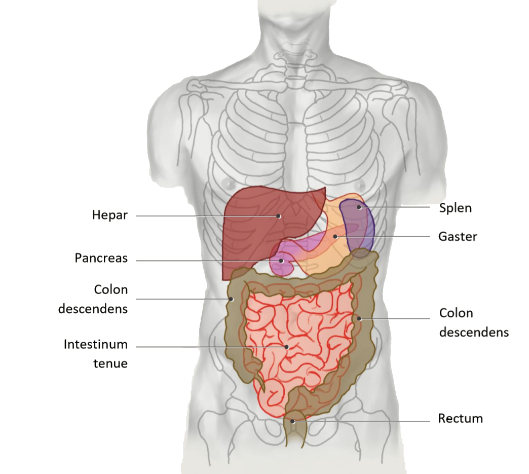
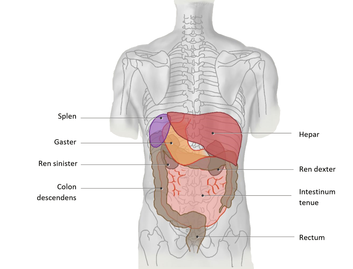
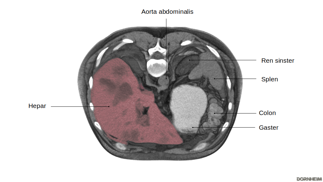
The liver is located in the right upper abdomen, but extends beyond the epigastrium into the left upper abdomen. It thereby pushes far in front of the stomach, which leaves an impression (impressio gastrica) at the back of the left liver lobe. The right lobe of the liver comes into close contact with the colon (right flexure), the small intestine (pars superior), the upper part of the right kidney and the right adrenal gland. Its impression sites are also named accordingly: Impressio colica, duodenalis, renalis and suprarenalis. The position of the liver is strongly dependent on respiration, as its underside is fused to the diaphragm. Age and posture are equally crucial in determining the position of the liver. The gallbladder is located dorsally between the left and right lobes of the liver.
Liver in situ
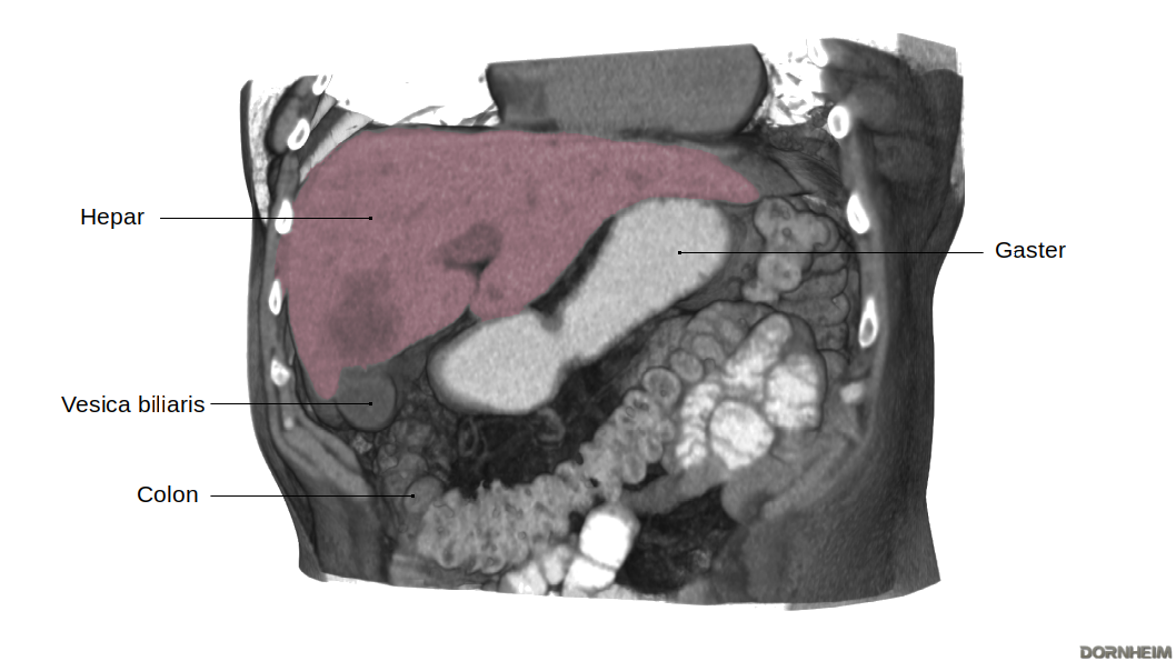
The thin area of tissue between the liver and the curvature of the stomach is called the omentum minus. This serous skin lines the peritoneal cavity and is the ventral border of the bursa omentalis. This is bordered on the right by the liver.
The downward-facing liver margin is sharp-edged and easily palpable in an enlarged liver. The liver extends from the right regio hypochondriaca, through the regio epigastrica, and into the left upper abdomen. The stomach can be seen at the left inferior border of the liver. The gallbladder lies on the underside of the liver and overhangs the inferior border of the liver.
Liver segments
The liver segments are formed by a total of 8 functional subunits of the liver. Each of these units has a branch of the portal vein, the bile duct and a segment branch of the arteria hepatica propria.
Assignment to Partes and Divisiones
| Pars hepatis sinistra | |
|---|---|
| Segment I | Pars posterior hepatis, Lobus caudatus |
|
Divisio lateralis sinistra |
| Segmentum mediale sinistrum (= Segmentum IV) subdivided into segment IVa and IVb |
Divisio medialis sinistra |
| Pars hepatis dextra | |
|---|---|
| Segmentum anterius mediale dextrum (= Segmentum V) Segmentum posterius mediale dextrum |
Divisio medialis dextra |
| Segmentum anterius laterale dextrum (= Segmentum VI) Segmentum posterius laterale dextrum |
Divisio lateralis dextra |
Since each subunit has its own supply route, the liver segments can be removed individually by surgery. The liver is still functional after the removal of a subunit, as are the removed subunits themselves. This makes a partial implantation of a liver possible.
Verschiedene Ansichten
| Sicht von dorsal auf die Pars superior der Facies diaphragmatica | Sicht von ventral auf die Facies diaphragmatica | Sicht von kaudal auf die Facies visceralis |
|
Der größte Teil der Leberoberfläche ist umgeben mit viszeralem Peritoneum. Jedoch bleibt die Area nuda als einzige peritonealfrei, ihre Oberfläche bildet die bindegewebige Kapsel. Außerhalb der Peritonealabdeckung verlassen die meist drei Vv. hepatica die Leber. Eine Besonderheit ist, dass bei der Leber nur die zuführende Arterie und zuführende V. portae hepatis als auch Ductus choledochus im Mesohepaticum verlaufen, die abführenden Venen hingegen nicht. An den Umschlagsorten des viszeralen in das parietale Peritoneum an der Unterseite des Zwerchfells, erscheint das bindegewebige Peritonealepithel als „Strang“ (Lig. coronarium). Aus dieser bindegewebigen Struktur entsteht am linken Leberlappen ein kleiner Zipfel (Appendix fibrosa hepatis). |
Aus der ventralen Ansicht sind die zwei Leberlappen, der große Lobus hepatis dexter und der kleinere Lobus hepatis sinister, gut zu erkennen. Das Lig. falciforme hepatis verläuft zwischen den beiden Lappen. Das Ligamentum falciforme hepatis bildet das Mesohepaticum ventrale und somit die Verbindung zwischen Leber und Bauchwand. |
Die kaudale Sicht ermöglicht einen Blick auf zwei weitere der insgesamt vier Leberlappen: den Lobus caudatus und den Lobus quadratus hepatis. Das Lig. hepatoduodenale dient der Leber zusammen mit dem Lig. hepatogastricum als Mesohepaticum dorsale und gehört topografisch zum Omentum minus. Die Gallenblase überragt den unteren Leberrand mit dem Fundus und liegt der Facies visceralis eng an. Der Hals der Gallenblase zeigt zur Leberpforte, wo sie in Kontakt mit den extrahepatischen Gallenwegen gelangt. |
Funktion
Die Leber hat als größtes und wichtigstes Stoffwechselorgan verschiedene Aufgaben im menschlichen Körper. Sie produziert lebenswichtige Proteine und ist maßgeblich an der Verwertung von Nahrungsbestandteilen sowie dem Abbau und der Ausscheidung von Stoffen beteiligt.
Erkrankungen
Freie Exploration
- Standard
- Fall: 1